
Uncategorized

breast ultrasound during breastfeeding

Breastfeeding & Breast Health: When Should a New Mother Consider a Breast Ultrasound?

Breastfeeding & Breast Health: When Should a New Mother Consider a Breast Ultrasound? Breastfeeding is a special phase of motherhood, but it also brings many physical changes. During lactation, the breasts naturally become fuller, denser, and more active as they produce milk. New mothers may occasionally notice tenderness, swelling, changes in breast texture, or even a lump. Although many breast changes during breastfeeding are benign, a new or persistent breast symptom should not simply be ignored because you are breastfeeding. A breast ultrasound during breastfeeding can be an important diagnostic tool when clinically recommended. It can help doctors evaluate a specific breast concern without using ionising radiation. At VCare Imaging & Diagnostic Centre in Kandivali, Mumbai, breast imaging services help women receive appropriate diagnostic evaluation based on their symptoms and their doctor’s or radiologist’s recommendations. Understanding what is normal during breastfeeding—and what deserves medical attention—can help new mothers make informed decisions about their breast health. Why Do Breasts Change During Breastfeeding? Pregnancy and breastfeeding cause significant hormonal and structural changes within breast tissue. Milk-producing glands become active, blood flow increases, and breasts may become larger and denser. These changes can sometimes make physical examination and breast imaging more complex. However, breastfeeding itself is not a reason to postpone evaluation of a concerning breast symptom. Medical imaging can be performed during lactation when clinically indicated. Some changes a breastfeeding mother may experience include: Many of these changes can have benign explanations. However, the cause of a persistent or unusual symptom cannot always be determined from symptoms alone. That is where appropriate clinical examination and, when recommended, breast imaging during breastfeeding can become important. What Is a Breast Ultrasound? A breast ultrasound, also known as breast sonography, uses high-frequency sound waves to create images of structures inside the breast. Unlike an X-ray, ultrasound does not use ionising radiation. During the examination, ultrasound gel is placed on the skin and a handheld transducer is moved over the area being examined. Images are produced in real time and interpreted by the appropriate medical professional. A breast ultrasound during breastfeeding may be used as part of the evaluation of a palpable lump, focal symptoms, or another breast concern, depending on the patient’s age, clinical findings and medical history. Current breast-imaging recommendations emphasize choosing imaging according to the specific clinical scenario rather than delaying investigation solely because a woman is lactating. When Should a Breastfeeding Mother Consider a Breast Ultrasound? Not every breastfeeding mother requires an ultrasound. Breast imaging is generally performed when there is a clinical reason or when recommended as part of an appropriate screening or diagnostic pathway. Here are some situations where a doctor may recommend further assessment. 1. A New or Persistent Breast Lump Finding a breast lump during breastfeeding can understandably cause anxiety. Lactation can be associated with benign conditions that may present as a lump. However, it is important not to assume that every lump is simply caused by breastfeeding. Current ACR guidance states that a new palpable abnormality during lactation should prompt appropriate imaging assessment. If you discover a lump that persists, changes, increases in size, or otherwise concerns you, consult your doctor rather than trying to diagnose it yourself. A breast ultrasound during breastfeeding may then form part of the diagnostic evaluation. 2. Persistent Localised Breast Pain Breast discomfort can occur during breastfeeding for several reasons. Feeding position, engorgement and other lactation-related issues may contribute to discomfort. However, persistent or focal breast pain deserves attention, particularly when accompanied by another symptom such as a lump, skin change, swelling or nipple discharge. A doctor can assess the symptoms and determine whether breast imaging is appropriate. 3. Swelling That Does Not Resolve Temporary fullness is common during breastfeeding. Persistent swelling—especially when confined to one area—should be discussed with a healthcare professional. Clinical evaluation helps determine whether the change appears related to breastfeeding or whether further assessment such as breast ultrasound is needed. 4. Persistent Signs of Infection or Inflammation Mastitis can occur during breastfeeding and may cause symptoms such as breast pain, redness, warmth and swelling, sometimes accompanied by fever or feeling unwell. Symptoms that do not improve as expected require medical review. Current ACR guidance specifically identifies unresolving infectious symptoms during lactation as a reason for imaging assessment. Ultrasound may be used in certain clinical situations to help assess whether there is a collection or another finding requiring further management. 5. Unusual Nipple Discharge Milk production is expected during breastfeeding, but a new or clinically suspicious nipple discharge should be evaluated rather than automatically attributed to lactation. The appropriate assessment depends on the nature of the discharge and other symptoms. ACR guidance includes clinically suspicious nipple discharge among the concerns that warrant imaging assessment during lactation. 6. Changes in the Skin or Nipple New breast changes should be discussed with a healthcare professional, particularly if they persist. These can include noticeable changes involving: These signs do not automatically indicate breast cancer, and there are many possible causes. The important point is that persistent or unexplained changes deserve proper evaluation. Is Breast Ultrasound Safe During Breastfeeding? Breast ultrasound does not involve ionising radiation and is widely used as a breast-imaging modality. The appropriate test, however, depends on factors including your symptoms, age, breast cancer risk and clinical examination. Women should therefore avoid assuming that ultrasound is always the only test needed simply because they are breastfeeding. Current evidence supports the use of ultrasound, mammography and, in selected circumstances, MRI for evaluating breast concerns in lactating patients. Can You Have a Mammogram While Breastfeeding? Yes. Mammography is not automatically contraindicated simply because you are breastfeeding. Current guidance supports mammography during lactation when it is appropriate for the patient’s age, risk or clinical situation. For example, the latest ACR criteria list mammography and digital breast tomosynthesis as usually appropriate for average-risk breast cancer screening in lactating women aged 40 or older. For diagnostic concerns, the imaging pathway may involve ultrasound, mammography, or both depending on age and the nature of the

Breastfeeding & Breast Health: When Should a New Mother Consider a Breast Ultrasound? Read More »

Best Time to Get a Mammography Test

Best Time to Get a Mammography Test: A Complete Guide to Early Breast Cancer Detection

Best Time to Get a Mammography Test: Everything You Need to Know Breast cancer is one of the most common cancers affecting women worldwide. The good news is that early detection through regular mammography screening significantly increases the chances of successful treatment. Many women often wonder, “What is the best time to get a mammography test?” Understanding the right age, timing, and preparation for this screening can help detect abnormalities before symptoms even appear. At VCare Imaging & Diagnosis Center, we believe that awareness and timely screening play a vital role in maintaining breast health. This comprehensive guide explains when you should schedule your mammography, factors that influence the timing, preparation tips, and frequently asked questions. What Is a Mammography Test? A mammography test is a specialized X-ray examination of the breast designed to detect early signs of breast cancer and other breast abnormalities. It can identify tiny lumps or calcium deposits that may not be noticeable during a physical examination. Mammograms are considered the gold standard for breast cancer screening because they can detect cancer years before it becomes physically noticeable. Why Is Mammography Important? Early-stage breast cancer often develops without any obvious symptoms. A routine mammogram can detect changes in breast tissue long before a lump becomes large enough to feel. Benefits of Mammography Early detection often means less invasive treatment and better long-term outcomes. Best Time to Get a Mammography Test The best time to get a mammography test depends on several factors, including your age, menstrual cycle, family history, and overall risk of breast cancer. If You Are Between 40 and 49 Years Women in this age group should discuss screening recommendations with their healthcare provider. Depending on individual risk factors, mammography may be recommended every one to two years. If You Are 50 Years and Above Most healthcare professionals recommend getting a mammogram every one to two years. Regular screening during this age group plays a crucial role in detecting breast cancer early. If You Have a Family History of Breast Cancer Women with a strong family history or genetic mutations such as BRCA1 or BRCA2 may need to begin mammography screening before the age of 40. Your doctor may also recommend breast MRI in addition to mammography. Best Time During Your Menstrual Cycle If you are menstruating, scheduling your mammogram at the right time can improve comfort and image quality. The ideal time is: Avoid scheduling your mammogram just before your period because breast tenderness can make the examination more uncomfortable. Signs That You Should Not Delay a Mammography Even if your routine screening is not yet due, consult your doctor immediately if you notice: Diagnostic mammography may be recommended instead of routine screening. How Often Should You Get a Mammogram? The screening interval depends on your age and health history. Age Group Recommended Frequency Under 40 Only if recommended by your doctor 40–49 Every 1–2 years (based on risk) 50–74 Every 1–2 years Above 75 As advised by your healthcare provider How to Prepare for a Mammography Test Preparing properly ensures accurate imaging results. Before Your Appointment These simple steps help improve image quality and reduce the need for repeat scans. What Happens During the Procedure? A mammography examination usually takes around 15 to 30 minutes. During the procedure: Compression may feel uncomfortable for a few seconds, but it is necessary to obtain clear images while minimizing radiation exposure. Is Mammography Painful? Most women describe mammography as mildly uncomfortable rather than painful. The amount of discomfort varies depending on: Scheduling your appointment after your menstrual period often makes the examination more comfortable. Can Mammography Detect Breast Cancer Early? Yes. Mammography can detect breast cancer before symptoms appear, often identifying tumors that are too small to be felt during a self-examination. Studies have consistently shown that regular mammography screening reduces deaths from breast cancer by enabling earlier diagnosis and treatment. Who Is at Higher Risk? Certain women may require earlier or more frequent screening. Risk factors include: If you have one or more of these risk factors, consult your healthcare provider about a personalized screening schedule. Benefits of Regular Mammography Screening Routine mammograms offer several long-term health benefits. Early Detection Detects cancer before symptoms develop. Better Treatment Outcomes Earlier diagnosis often leads to less invasive treatment options and improved survival rates. Reduced Healthcare Costs Treating cancer at an early stage is generally less complex and less expensive than treating advanced disease. Improved Peace of Mind Regular screening helps women stay proactive about their breast health. Common Myths About Mammography Myth 1: Mammograms Cause Cancer False. Mammography uses a very low dose of radiation, and the benefits of early detection far outweigh the minimal risk. Myth 2: Only Women with Symptoms Need Mammograms False. Many breast cancers have no symptoms in their early stages, which is why routine screening is essential. Myth 3: Breast Self-Exams Replace Mammography False. Breast self-examinations are helpful for becoming familiar with your breasts, but they do not replace mammography. Imaging can detect abnormalities long before they can be felt. Frequently Asked Questions Why Choose VCare Imaging & Diagnosis Center? At VCare Imaging & Diagnosis Center, we are committed to providing accurate, comfortable, and patient-focused diagnostic services. Our advantages include: Our goal is to help patients receive accurate diagnoses while ensuring a smooth and reassuring experience. Final Thoughts Knowing the best time to get a mammography test is an important step toward protecting your breast health. Whether you are scheduling your first screening or maintaining a regular screening routine, timely mammography can help detect breast cancer at its earliest and most treatable stage. If you are over 40, have a family history of breast cancer, or notice any unusual breast changes, don’t delay your screening. Early detection saves lives, and regular mammograms remain one of the most effective tools for maintaining long-term breast health. Book your mammography appointment at VCare Imaging & Diagnosis Center today and take a proactive step toward early breast cancer detection and better health. https://maps.app.goo.gl/m8uoy6bBcuibEtkYA

Best Time to Get a Mammography Test: A Complete Guide to Early Breast Cancer Detection Read More »

Best 5 Diagnostic Centers in Kandivali (2026)

Best 5 Diagnostic Centers in Kandivali (2026) Finding the best diagnostic center in Kandivali is one of the most important decisions you can make for your health. Whether your doctor has recommended an MRI scan, CT scan, ultrasound, blood test, mammography, or a complete preventive health check-up, the quality of the diagnostic centre directly impacts the accuracy of your diagnosis and, ultimately, your treatment. A diagnostic test is more than just a report—it is the foundation of medical decision-making. Accurate imaging and pathology results help doctors identify diseases early, monitor existing conditions, and recommend the most effective treatment plan. Even the most experienced physician relies on high-quality diagnostic reports to provide the best possible care. Kandivali has several diagnostic centres offering imaging and laboratory services. However, the quality of technology, radiologists, reporting standards, hygiene, and patient care can vary significantly from one centre to another. Choosing a trusted diagnostic centre ensures you receive accurate results, timely reports, and a comfortable experience. To help you make an informed decision, we’ve compiled a list of some of the best diagnostic centers in Kandivali based on the range of services, advanced technology, patient experience, qualified specialists, and commitment to quality healthcare. Why Choosing the Right Diagnostic Centre Matters Many people select a diagnostic centre simply because it is close to home or offers low prices. While convenience and affordability are important, they should never come at the expense of diagnostic accuracy. A reliable diagnostic centre offers much more than routine testing. It combines experienced radiologists, modern imaging equipment, standardized protocols, skilled technicians, and quality control measures to ensure every report is accurate and clinically useful. Here are some key factors to consider when choosing a diagnostic centre: 1. Advanced Diagnostic Technology Modern equipment provides clearer images, faster scanning times, and greater diagnostic precision. Technologies such as Digital MRI, Multi-Slice CT Scan, Digital Mammography, 3D/4D Ultrasound, Colour Doppler, and Digital X-Ray improve image quality while enhancing patient comfort. 2. Experienced Radiologists Medical imaging requires expert interpretation. Qualified radiologists play a vital role in identifying abnormalities accurately, ensuring that reports are reliable and clinically meaningful. 3. Comprehensive Services Under One Roof A diagnostic centre that offers MRI, CT Scan, Ultrasound, Pathology, ECG, 2D Echo, Mammography, Bone Density Scan, and preventive health packages saves patients time and simplifies the diagnostic process. 4. Quality Standards Centres that follow recognized quality protocols and maintain high standards of hygiene, equipment maintenance, and reporting are generally more dependable. 5. Timely Reporting Quick and accurate reports help doctors begin treatment sooner, especially in emergency situations where time is critical. 6. Patient-Centric Care Friendly staff, clean facilities, efficient appointment scheduling, and clear communication all contribute to a better healthcare experience. 1. VCare Imaging & Diagnosis Center – Our Featured Recommendation When comparing the best diagnostic centers in Kandivali, VCare Imaging & Diagnosis Center stands out as a comprehensive destination for advanced imaging, pathology, and preventive healthcare services. With its focus on diagnostic excellence, modern technology, and patient-first approach, VCare has earned the trust of patients and referring doctors alike. Whether you need a routine blood test or an advanced MRI scan in Kandivali, VCare offers a wide range of diagnostic services under one roof, making healthcare more convenient and efficient. Comprehensive Diagnostic Services VCare provides an extensive range of diagnostic solutions, including: This comprehensive approach allows patients to complete multiple investigations at a single location, saving valuable time and reducing the need to visit multiple facilities. Advanced Imaging Technology Accurate diagnosis begins with high-quality imaging. VCare uses advanced diagnostic equipment designed to deliver detailed images that support precise medical evaluation. High-resolution imaging enables doctors to identify conditions affecting the brain, spine, joints, heart, lungs, abdomen, bones, and other organs with greater confidence. Modern technology also helps improve patient comfort by reducing scan times and producing clearer diagnostic images. Experienced Radiologists and Skilled Professionals Behind every accurate diagnostic report is an experienced team of radiologists and trained technicians. VCare’s medical professionals follow standardized imaging protocols and work closely with referring physicians to ensure every report provides meaningful clinical information. Their expertise plays a vital role in detecting diseases at an early stage and assisting doctors in making informed treatment decisions. Focus on Preventive Healthcare Healthcare today is not only about treating diseases-it is also about preventing them. VCare encourages regular preventive health screenings to identify potential health concerns before symptoms appear. Preventive health packages help monitor: Early diagnosis often leads to better treatment outcomes and lower healthcare costs. Comfortable Patient Experience Many patients feel anxious before undergoing MRI scans or other imaging procedures. VCare strives to create a welcoming environment where patients feel informed, supported, and comfortable throughout their visit. From appointment scheduling to report collection, the focus remains on delivering a smooth and hassle-free experience. Patients also appreciate: Why We Recommend VCare Imaging & Diagnosis Center While several diagnostic centres serve Kandivali, VCare stands out because of its combination of comprehensive services, advanced technology, experienced professionals, and patient-focused care. Key strengths include: For patients seeking reliable diagnostics, VCare offers a balanced combination of quality, convenience, and comprehensive healthcare services. Services Available at VCare Imaging & Diagnosis Center Who Should Visit VCare? VCare serves a wide range of patients, including: 2. Dr. Vyas Diagnostic Centre – A Well-Known Local Diagnostic Facility Dr. Vyas Diagnostic Centre is one of the established diagnostic centres serving patients in and around Kandivali. The centre offers a range of routine diagnostic services, including pathology tests, ultrasound, X-ray, and general health investigations. Many patients visit this centre for routine laboratory tests and basic diagnostic requirements. It caters to individuals looking for common health screenings and physician-recommended investigations. Key Services Dr. Vyas Diagnostic Centre is suitable for routine diagnostics. However, patients requiring advanced imaging modalities such as MRI, CT Scan, Digital Mammography, CBCT, or DEXA Scan may need to consider a centre with a broader range of diagnostic services. 3. Siddhi Diagnostic Centre Siddhi Diagnostic Centre has been serving local residents by providing pathology services and basic imaging investigations.

Best 5 Diagnostic Centers in Kandivali (2026) Read More »

Mammography After Age 40

Why Mammography is Important After Age 40

Why Mammography After Age 40 is Essential for Women’s Health As women age, regular health screenings become increasingly important, especially for breast health. One of the most recommended diagnostic tests for women above 40 is a mammography test. Doctors worldwide advise women to begin routine mammography after age 40 because early detection of breast abnormalities can significantly improve treatment outcomes and survival rates. Breast cancer is one of the most common cancers among women, but with timely screening and advanced diagnostic imaging, it can often be detected at an early stage. At Vcare Imaging & Diagnostic Centre, advanced digital mammography services help women take proactive steps toward better health and early diagnosis. What is Mammography? Mammography is a specialized breast imaging test that uses low-dose X-rays to detect abnormalities in breast tissue. It helps identify small lumps, calcifications, or signs of breast cancer even before symptoms appear. There are mainly two types of mammography: Modern digital mammography provides clearer images and improves the accuracy of breast cancer screening. Why Mammography After Age 40 is Important 1. Early Detection of Breast Cancer The biggest advantage of mammography after age 40 is early detection. Breast cancer often develops silently without noticeable symptoms in the initial stages. Mammograms can detect cancer years before it becomes physically noticeable. Early diagnosis can: 2. Breast Cancer Risk Increases with Age As women grow older, the risk of developing breast cancer gradually increases. Women above 40 are more likely to develop breast tissue changes that require medical evaluation. Regular breast cancer screening helps monitor these changes and ensures timely medical attention if needed. 3. Mammography Can Detect Tiny Abnormalities A mammography test can identify: These abnormalities are often too small to be felt during a physical examination. 4. Helps in Preventive Healthcare Preventive healthcare focuses on detecting health conditions before symptoms worsen. Routine mammography after age 40 is considered an essential preventive health screening for women. Doctors usually recommend: Who Should Get a Mammography Test? A mammography test is recommended for: Consulting a radiologist or healthcare professional can help determine the right screening schedule. Common Symptoms That Should Not Be Ignored Even though routine screening is important, women should immediately consult a doctor if they notice: A timely breast imaging test can help identify the exact cause. How to Prepare for a Mammography Test Preparing for a mammography test is simple. Here are a few useful tips: The procedure usually takes around 15–20 minutes. Is Mammography Painful? Many women worry about discomfort during the test. Mammography may cause mild pressure for a few seconds while images are captured, but the procedure is generally quick and tolerable. The benefits of early detection far outweigh temporary discomfort. Benefits of Digital Mammography Modern digital mammography offers several advantages over traditional systems: Digital imaging technology plays an important role in accurate breast cancer diagnosis. Why Choose Vcare Imaging & Diagnostic Centre for Mammography in Mumbai? Choosing the right diagnostic centre is essential for accurate reporting and patient comfort. Vcare Imaging & Diagnostic Centre offers advanced diagnostic imaging services with experienced radiologists and modern technology. Highlights: Women looking for a reliable mammography test in Mumbai can benefit from professional diagnostic support and quality imaging services. Conclusion Regular mammography after age 40 is one of the most effective ways to protect women’s breast health. Early detection through routine screening can save lives, reduce treatment complications, and improve long-term health outcomes. Women should never ignore regular breast cancer screening and preventive health checkups. With advanced imaging technology and expert diagnostic care, early diagnosis becomes easier and more accurate. If you are planning a mammography test in Mumbai, consult experienced professionals at Vcare Imaging & Diagnostic Centre for reliable and advanced breast imaging services.

Why Mammography is Important After Age 40 Read More »

Early Breast Cancer Symptoms

Early Breast Cancer Symptoms Every Woman Should Never Ignore

Learn the early breast cancer symptoms every woman should never ignore. Understand the importance of breast cancer screening and mammography test for early detection at the best imaging and diagnostic center in Kandivali. Breast cancer has become one of the most common health concerns affecting women globally. Despite advancements in medical science, many cases are still diagnosed in later stages simply because the early warning signs were ignored or overlooked. This is why awareness about early breast cancer symptoms is extremely important for every woman. Early detection plays a crucial role in improving survival rates and ensuring successful treatment outcomes. When breast cancer is identified at an early stage, treatment becomes less complicated and the chances of recovery increase significantly. Regular breast cancer screening and diagnostic tests such as a mammography test help detect abnormalities before they become serious health concerns. Unfortunately, many women ignore small changes in their bodies because they assume the symptoms are temporary or harmless. However, even minor changes in breast tissue can sometimes indicate underlying health issues that require medical attention. For accurate diagnosis and preventive screening, visiting the best imaging and diagnostic center in Kandivali like Vcare Imaging & Diagnostic Centre can help ensure reliable reports, advanced imaging technology, and professional healthcare support. Understanding Breast Cancer Early detection is one of the most effective ways to fight breast cancer. When the disease is diagnosed in its early stages, treatment is usually more successful and less invasive. Here are some important benefits of early breast cancer detection: Diagnostic tools such as the mammography test allow doctors to identify abnormal changes in breast tissue even before physical symptoms become noticeable. Early Breast Cancer Symptoms Every Woman Should Watch For Recognizing early breast cancer symptoms is essential for timely medical evaluation. While not every symptom indicates cancer, any unusual change should be checked by a healthcare professional. Lump in the Breast or Underarm One of the most common early breast cancer symptoms is a lump in the breast or underarm area. These lumps are often painless and may feel hard or irregular in shape. Although many breast lumps are benign, any new lump should be examined through proper medical evaluation and imaging tests. Changes in Breast Size or Shape A noticeable change in the size or shape of the breast may sometimes indicate underlying abnormalities. If one breast suddenly appears swollen, enlarged, or differently shaped compared to the other, it is advisable to consult a healthcare professional and consider breast cancer screening. Skin Changes on the Breast Another important sign to watch for includes unusual changes in the skin of the breast. These may include: Such changes may indicate underlying breast tissue abnormalities and should be evaluated promptly. Nipple Changes or Inversion Sudden nipple changes may also be among the early breast cancer symptoms. These changes can include: These symptoms should never be ignored and should be evaluated by a healthcare professional. Unusual Nipple Discharge Unexpected discharge from the nipple without squeezing or breastfeeding can sometimes signal a problem. Although nipple discharge can occur due to non-cancerous conditions, bloody or clear discharge should be investigated through medical examination and diagnostic tests like a mammography test. Persistent Breast Pain Breast pain is commonly associated with hormonal changes or menstrual cycles. However, persistent pain in a specific area of the breast should not be ignored. If pain occurs along with swelling, lumps, or skin changes, medical evaluation is necessary. Importance of Mammography in Breast Cancer Screening A mammography test is one of the most reliable diagnostic tools used in breast cancer screening. It uses low-dose X-rays to capture detailed images of breast tissue and detect abnormalities that cannot be felt during a physical examination. H3: Benefits of a Mammography Test Doctors generally recommend that women above the age of 40 undergo regular mammography screening. Women with a family history of breast cancer may need to start screening earlier. Who Should Get Breast Cancer Screening? Breast cancer screening is important for women of different age groups depending on their health history and risk factors. Women who should consider regular screening include: Regular screening helps detect abnormalities early and ensures timely treatment. Why Choosing the Right Diagnostic Centre Matters When it comes to early detection, choosing the right diagnostic centre plays a vital role. Accurate imaging, advanced equipment, and experienced professionals ensure reliable diagnostic results. For individuals looking for the best imaging and diagnostic center in Kandivali, Vcare Imaging & Diagnostic Centre offers advanced imaging services and professional diagnostic care. The center focuses on delivering accurate reports, maintaining high medical standards, and providing a comfortable environment for patients undergoing diagnostic tests Conclusion Awareness about early breast cancer symptoms can help women detect potential health concerns before they become serious. Paying attention to changes in the body, performing regular self-examinations, and undergoing routine breast cancer screening are important steps in maintaining breast health. Diagnostic tests such as a mammography test play a crucial role in identifying abnormalities at an early stage. Early detection not only increases the chances of successful treatment but also helps reduce complications and improve overall health outcomes. Women should prioritize their health by scheduling regular screenings and consulting healthcare professionals whenever unusual symptoms appear. Choosing a reliable healthcare facility like the best imaging and diagnostic center in Kandivali ensures accurate diagnosis and timely medical support. Taking preventive action today can make a significant difference in protecting your health and well-being in the future.

Early Breast Cancer Symptoms Every Woman Should Never Ignore Read More »

Best Imaging and Diagnostic Center in Kandivali Mumbai

What is the difference between a pathology lab and a diagnostic centre?

In today’s fast-paced healthcare environment, early and accurate diagnosis plays a pivotal role in ensuring effective treatment. However, with an ever-expanding landscape of medical facilities, it’s not uncommon for patients to feel confused by the terminology. One such point of confusion arises when distinguishing between a pathology lab and a diagnostic centre. Though both contribute significantly to the diagnostic process, they serve distinct roles within the healthcare system. For those looking for a reliable pathology lab in Kandivali or a comprehensive diagnostic centre in the same locality, understanding these differences can help you make informed choices based on your specific health needs. Let’s explore the unique roles, capabilities, and purposes of pathology labs and diagnostic centres in detail. Understanding a Pathology Lab A pathology lab, also known as a clinical or medical laboratory, plays a critical role in diagnosing various conditions by examining biological samples. These samples can range from blood and urine to tissue and stool. What sets pathology labs apart is their reliance on scientific methodologies and highly trained professionals who analyse these specimens to detect diseases at the microscopic or even molecular level. A pathology lab primarily focuses on diagnosing diseases through the study of samples such as blood, urine, tissues, or other bodily fluids. These labs operate at a cellular and molecular level, enabling clinicians to gain insights into what’s happening within the body, even before symptoms appear. For instance, when a doctor requests a blood test, a urine test, or a biopsy analysis, it is the pathology lab that performs and interprets these tests. There are several subdivisions within a pathology lab: Clinical Pathology involves testing body fluids to detect infections, diabetes, liver disorders, and more. This includes routine blood tests like CBC (Complete Blood Count), lipid profiles, and liver function tests. Common Tests Conducted in Pathology Labs Tests conducted in a pathology lab are both routine and highly specialised. These include: Each of these tests is performed with strict protocols to ensure accuracy. At a trusted pathology lab in Kandivali, patients can expect precision, confidentiality, and quick turnaround times. When and why are pathology tests prescribed? Doctors usually prescribe pathology tests when they need to: Common scenarios include checking for infections, tracking chronic illnesses like diabetes or thyroid disorders, or screening for deficiencies like anaemia or vitamin D. What happens during a pathology test? When you visit a pathology lab, a trained technician or phlebotomist collects the necessary samples, which might include blood, urine, or swabs. These are carefully labelled and sent for analysis. Depending on the test, it might be processed by machines or manually examined under a microscope. Results are usually delivered within a few hours to a couple of days. A reputable pathology lab ensures hygiene, safety, and timely reporting. What is a diagnostic centre? A diagnostic centre offers a broader range of services than a pathology lab. While it includes pathology services, it also provides imaging tests such as X-rays, MRIs, CT scans, and ultrasounds. In essence, a diagnostic centre is a one-stop solution for multiple diagnostic needs. For individuals in need of a full-body health screening or imaging tests, a diagnostic centre in Kandivali is an ideal choice. Key tests conducted in diagnostic centres Diagnostic centres offer a wide array of tests, including: Blood and urine tests (from the pathology lab wing) These centres are equipped with advanced machinery and skilled radiologists and technicians to ensure precise results. Many diagnostic centres also offer health packages tailored to different age groups and medical conditions. Purpose of a Diagnostic Centre Diagnostic centres aim to offer a holistic view of a patient’s health. By combining pathology and imaging, they make it easier for doctors to detect diseases early, plan treatments more effectively, and monitor recovery over time. From walk-in patients to referrals, these centres accommodate a wide range of healthcare needs under one roof. What to expect during a visit to a diagnostic centre? At a well-established diagnostic centre, the patient journey is designed to be smooth and hassle-free. Upon arrival, patients are registered and guided through their tests. Imaging tests may require specific preparations, such as fasting or drinking fluids. Staff members explain each step clearly. After tests, results are processed and usually delivered electronically or as physical reports. Some centres even offer consultations with in-house doctors based on the test outcomes. Which one should you choose? Your choice should depend on the tests recommended by your doctor. If you only need a blood or urine test, a specialised pathology lab in Kandivali is sufficient. However, if your health check-up involves multiple screenings, such as scans and pathology tests, a diagnostic centre is a better option. Conclusion Understanding the difference between a pathology lab and a diagnostic centre empowers you to make informed healthcare decisions. Whether you need routine blood tests or more complex imaging, both facilities play essential roles in identifying and managing health issues. At VCare Imaging & Diagnostic Centre in Kandivali, patients benefit from the convenience of having both services under one roof. From MRI and CT scans to comprehensive blood work, VCare offers precise, timely, and trustworthy diagnostics to ensure your health remains a top priority. Your Trusted Diagnostic Centre in Kandivali Discover top-quality healthcare services at VCare Imaging & Diagnostic Centre. We offer a wide range of services, including Sonography, Mammography, MRI, CT Scan, and advanced Blood Tests. Whether you need a specialised pathology lab in Kandivali or a full-service diagnostic centre, VCare is your destination for reliable and compassionate healthcare.

What is the difference between a pathology lab and a diagnostic centre? Read More »

Best Imaging and Diagnostic Center in Kandivali Mumbai

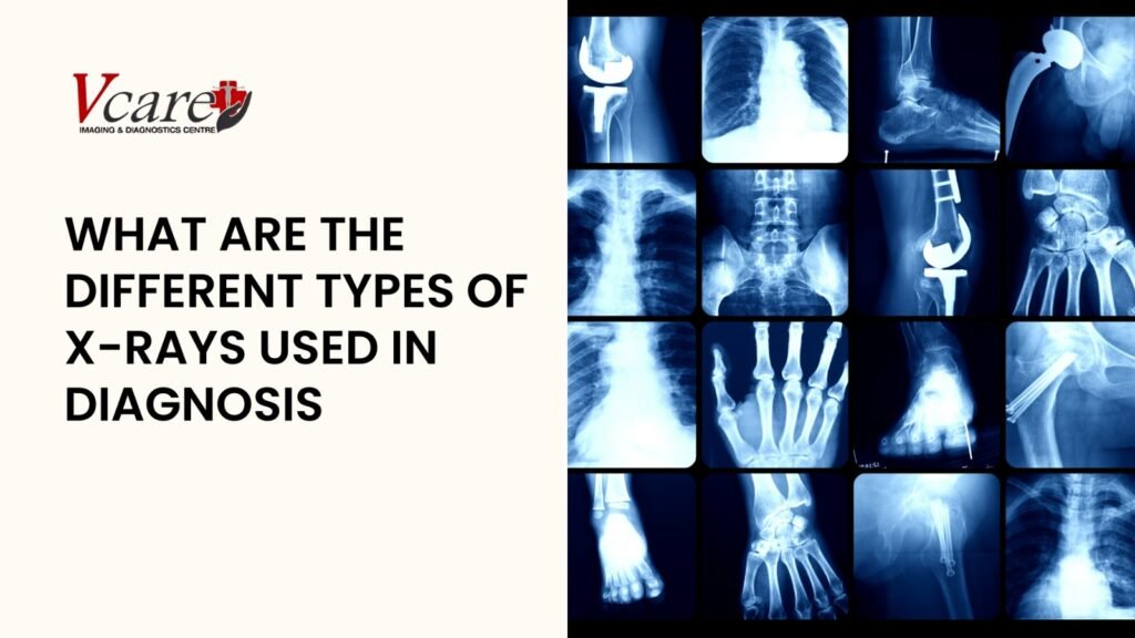
What are the different types of X-rays used in diagnosis?

Introduction X-rays have revolutionised the way medical professionals diagnose, monitor, and manage a wide range of health conditions. Discovered by Wilhelm Conrad Roentgen in 1895, X-rays use electromagnetic radiation to create images of the inside of the body, offering a non-invasive, quick, and reliable diagnostic method. Today, they form the backbone of imaging services in most diagnostic centres in Kandivali and across the globe. Whether it’s detecting bone fractures, spotting infections, evaluating lung conditions, or guiding surgical procedures, X-rays play a pivotal role in modern medicine. Depending on the diagnostic need, different types of X-rays are employed, each tailored to highlight specific tissues, organs, or abnormalities. From the standard chest X-ray to advanced techniques like fluoroscopy and computed tomography (CT), the applications are vast and continually evolving with technological advancements. If you’re visiting an x-ray centre in Kandivali or a diagnosis lab in Kandivali, understanding the type of X-ray recommended can help ease anxiety and offer clarity on what your doctor is looking for. In this article, we’ll walk you through the major types of X-rays used in diagnosis and how each serves a unique purpose in identifying and treating medical conditions. What is an X-Ray? An X-ray is a form of electromagnetic radiation that can pass through most objects, including the human body. When X-rays are directed through your body, they are absorbed in different amounts depending on the density of the material they pass through. Dense materials like bones absorb more X-rays and appear white on the X-ray image, whereas softer tissues absorb less and appear in shades of grey or black. X-rays allow doctors to view internal structures without making a single incision. That’s why they’re considered essential tools in nearly every diagnostic centre in Kandivali and play a major role in accurate diagnosis and timely treatment. Whether it’s a simple bone fracture or a deeper internal issue, X-rays offer a glimpse into areas the naked eye can’t see. How do X-Rays work? When you go in for an X-ray at a trusted x-ray centre in Kandivali, the machine emits a controlled amount of radiation that passes through the targeted area of your body. As the rays travel through, they are captured by a detector or film placed on the opposite side. Different tissues absorb radiation differently, bones block the rays more and appear white; air-filled spaces like the lungs appear black; muscles and fat show up in varying shades of grey. This contrast creates a detailed image of your internal structure that doctors can use to identify abnormalities, injuries, or diseases. Today, most diagnosis labs in Kandivali use digital X-ray technology, which allows images to be stored, shared, and enhanced electronically for faster, more accurate diagnoses. Types of X-Rays Different medical conditions require different types of X-rays. Let’s explore the most commonly used ones you might come across at a diagnostic centre in Kandivali. The Role of X-Rays in Diagnosis X-rays serve as the first line of imaging in countless medical scenarios. If you’re visiting a diagnosis lab in Kandivali, there’s a high chance that the first step your doctor might suggest is an X-ray to get a basic understanding of the condition. For injuries, infections, unexplained pain, or routine screenings, X-rays are universally relied upon. They help doctors confirm clinical suspicions, assess the severity of a condition, plan treatment strategies, and even guide procedures like the insertion of catheters or pacemakers. X-rays offer clarity and accuracy, especially when time is of the essence. When should you get an X-Ray? While only a doctor can determine when you need an X-ray, some common indications include persistent pain, visible swelling, difficulty in movement, or injury due to an accident. Routine screenings such as mammograms or dental checks also involve X-rays even in the absence of symptoms. You may be advised to undergo an X-ray if your healthcare provider suspects internal abnormalities that can’t be diagnosed externally. Regular scans are also common for those undergoing treatment for chronic illnesses, to monitor progress or catch any complications early. Advantages of Using X-Rays One of the main reasons X-rays are so widely used in every x-ray centre in Kandivali is because of their efficiency. They are quick, painless, and often provide immediate results. For patients, this means less waiting and quicker diagnosis. They are non-invasive, making them safer and easier for people of all ages, including children and elderly patients. Compared to other imaging methods like MRI or CT, basic X-rays are more affordable and accessible, ensuring more people can benefit from early diagnosis and intervention. Digital X-rays also reduce radiation exposure and provide enhanced image quality, helping doctors make better-informed decisions. The Importance of X-Rays in Early Cancer Detection X-rays are not just diagnostic, they’re life-saving. One of their most vital roles is in the early detection of cancer. Mammograms, for example, are responsible for saving countless lives by identifying breast cancer before symptoms appear. Chest X-rays can detect tumours, lung nodules, or pleural effusions early, allowing for quicker treatment. At a well-equipped diagnostic centre in Kandivali, regular screening using X-ray technology is part of preventive healthcare, especially for individuals at high risk. Early detection is often the difference between a simple treatment and a prolonged battle. X-rays help bridge that gap with speed and precision. How to prepare for an X-Ray? Preparation for an X-ray is usually minimal, but it does depend on the type of test. At most x-ray centres, you’ll be asked to remove any metal objects like jewellery or belts. In some cases, you may need to change into a hospital gown. If the X-ray involves the stomach or intestines, you might be asked to fast for a few hours prior. For women, it’s essential to inform the technician if there’s any chance of pregnancy. The actual procedure takes only a few minutes and is completely painless. The technicians at a good diagnostic lab will guide you through every step, ensuring comfort and clarity. Conclusion In the ever-evolving world of healthcare, X-rays remain a cornerstone in accurate medical diagnosis. From identifying fractures and infections to detecting early signs of cancer, X-rays continue

What are the different types of X-rays used in diagnosis? Read More »

Best Imaging and Diagnostic Center in Kandivali Mumbai

Optimizing Heart Health: The Role of Cholesterol and Lipid Profile Tests

Introduction Heart disease remains one of the leading causes of death worldwide and elevated cholesterol is a key contributor. According to global statistics, nearly 39% of adults have raised total cholesterol levels, significantly increasing their risk of cardiovascular disease (CVD). Poor dietary habits, lack of physical activity, smoking, and genetic factors all influence cholesterol levels. Monitoring your lipid profile is not just about numbers, it’s a proactive step toward preventing heart attacks and strokes. Understanding your cholesterol levels and managing them early can save lives and improve long-term health outcomes. If you reside in Mumbai, finding the best diagnostic centre for cholesterol test in Kandivali ensures timely and accurate monitoring of your cardiovascular health. What is Cholesterol? Cholesterol is a waxy, fat-like substance that your body needs to function properly. It plays an essential role in producing hormones, vitamin D, and substances that help digest food. While your body naturally produces cholesterol, it also comes from dietary sources, especially animal-based products. However, when cholesterol levels are too high — particularly LDL (bad cholesterol) it can build up in your arteries and increase the risk of heart disease and stroke. That’s why maintaining a balance between HDL (good cholesterol) and LDL is critical for cardiovascular health. Types of Cholesterol: Understanding the Lipid Profile A lipid profile is a comprehensive blood test that gives detailed insights into your cholesterol status. It includes the following key components: For residents of Mumbai, especially those looking for a lipid profile test in Kandivali, a reliable diagnostic centre will measure all these components to provide an accurate risk assessment. Why is Lipid Profile Testing important? A cholesterol test in Kandivali can offer early detection of elevated lipid levels, enabling prompt intervention. Whether you’re undergoing preventive screening or monitoring existing heart issues, lipid tests help evaluate treatment efficacy and provide actionable insights into your heart health. Cardiovascular diseases often develop silently, and lipid abnormalities are one of the earliest indicators of risk. Values may vary based on age, gender, and underlying health conditions. For accurate results, choose the best diagnostic centre for cholesterol test in Kandivali to ensure consistency and reliability. What to Expect from a Lipid Profile Test? A lipid panel test in Kandivali involves a simple blood draw, usually after fasting for 9-12 hours to get accurate triglyceride readings. The process is safe, quick, and typically takes only a few minutes. Diagnostic centres like those in Kandivali will guide you through the pre-test preparations and post-test interpretations, making it a smooth and informative process. Risk Factors for High Cholesterol Cholesterol levels are influenced by various factors: Getting a heart health checkup Kandivali is crucial for individuals with these risk factors, as it allows for timely management and prevention. How to Maintain Healthy Cholesterol Levels? Managing cholesterol involves lifestyle modifications and sometimes medication. Incorporate the following practices: Top cholesterol-lowering foods include nuts, seeds, olive oil, berries, legumes, and avocados. For individuals requiring medical guidance, visiting a cholesterol test in the Kandivali facility helps tailor a personalized plan. When should you get tested? For women post-menopause, individuals with diabetes, and those with a family history of heart disease, it is strongly advised to schedule a lipid profile test in Kandivali as part of regular health screenings. Conclusion In today’s fast-paced world, taking control of your health is more important than ever. Cardiovascular issues can creep in silently, but regular cholesterol and lipid testing provides an early warning system. Whether you’re in your 20s or your 60s, proactive testing and healthy habits go a long way. If you’re searching for the best diagnostic centre for cholesterol test in Kandivali, choose a reputable facility that offers detailed analysis, expert consultation, and prompt services. A heart health checkup in Kandivali is more than a routine exam, it’s a life-saving decision. Book your lipid panel test in Kandivali today and take the first step toward a heart-healthy future. Your heart will thank you.

Optimizing Heart Health: The Role of Cholesterol and Lipid Profile Tests Read More »

Best Imaging and Diagnostic Center in Kandivali Mumbai

Imaging Tests During Pregnancy: What’s Safe and What’s Not

Introduction Motherhood is a precious journey filled with both excitement and concerns. Pregnancy is a transformative experience that brings significant changes to a woman’s body, making maternal and fetal health a top priority. Imaging tests play a crucial role in monitoring fetal development and detecting potential complications. However, expecting mothers and their families often worry about the safety of these tests. Understanding the risks, benefits, and precautions of imaging techniques such as ultrasounds, MRIs, X-rays, and CT scans at a CT scan centre in Kandivali can help make informed healthcare decisions. Why Should One Do Imaging Tests During Pregnancy? Monitoring Fetal Development Imaging tests track the baby’s growth and development throughout pregnancy. Routine ultrasounds, including 3D and 4D sonography in Kandivali, allow healthcare providers to assess the baby’s size, movements, and overall health. These tests also help detect congenital anomalies early, ensuring timely medical intervention. Checking Fetal Position As pregnancy progresses, determining the baby’s position becomes crucial, especially before delivery. Ultrasounds help identify whether the baby is in a breech or transverse position, allowing doctors to plan necessary medical or manual interventions for a safe delivery. Assessing the Placenta and Amniotic Fluid The placenta and amniotic fluid are essential for fetal development. Imaging tests help evaluate placental function and detect conditions like placenta previa or placental insufficiency. Doctors also assess amniotic fluid levels to prevent complications such as oligohydramnios (low fluid) or polyhydramnios (excess fluid). Diagnosing High-Risk Pregnancy Complications Women with high-risk pregnancies, including those with gestational diabetes, preeclampsia, or multiple pregnancies, require frequent imaging tests. Advanced imaging techniques help diagnose maternal and fetal conditions without exposing the baby to unnecessary risks. Diagnosing Maternal Health Issues Imaging tests may be needed to assess maternal health conditions that could impact pregnancy. For example, they help diagnose kidney stones, appendicitis, or other complications requiring medical attention. Guiding Procedures Some imaging techniques assist doctors in performing procedures such as amniocentesis or chorionic villus sampling (CVS). These tests help diagnose genetic disorders and other fetal abnormalities, ensuring appropriate medical guidance for expecting parents. Precautions Before Undergoing Imaging Tests During Pregnancy When undergoing imaging tests during pregnancy, safety is a priority. Always inform your doctor and radiologist about your pregnancy before any scan. Here are essential precautions to follow: Safe Imaging Tests Ultrasound (2D, 3D, 4D) Ultrasound is considered completely safe during pregnancy because it relies on high-frequency sound waves rather than ionizing radiation. 3D and 4D sonography in Kandivali is widely used to assess fetal growth, organ development, and overall well-being without posing any known risks. MRI (Magnetic Resonance Imaging) MRI is also considered safe for pregnant women as it does not involve ionizing radiation. It provides detailed imaging of fetal structures and is often recommended when more precise imaging is needed. For those seeking advanced imaging, visiting an MRI centre in Kandivali ensures access to high-quality diagnostics with expert radiologists. Use with Caution X-rays X-rays should generally be avoided during pregnancy unless absolutely necessary. However, if needed for diagnosing fractures or dental issues, visiting an X-ray centre in Kandivali that follows strict safety protocols ensures minimal fetal radiation exposure. CT Scans CT scans use ionizing radiation and should only be performed if essential for diagnosing maternal health concerns. If required, selecting a trusted CT scan centre in Kandivali ensures that the procedure is performed with the lowest radiation dose and highest safety standards. Avoid Common Imaging Tests During Pregnancy Conclusion Pregnancy is a special time, and ensuring the safety of both mother and baby is essential. While some imaging tests raise concerns, advancements in medical technology have made most procedures safe when conducted under expert guidance. At V Care Diagnostics, we prioritize the well-being of expecting mothers by offering state-of-the-art imaging services, including 3D and 4D sonography in Kandivali, MRI, X-rays, and CT scans. Our experienced radiologists and advanced technology ensure accurate diagnoses while adhering to the highest safety standards. Book your imaging test today at our CT scan centre in Kandivali and ensure a safe and informed pregnancy experience.

Imaging Tests During Pregnancy: What’s Safe and What’s Not Read More »

Best Imaging and Diagnostic Center in Kandivali Mumbai

Why You Might Need a Pelvic Ultrasound and What It Can Reveal?

Introdcution Ever wondered what’s going on inside your body but wished there was a way to peek without all the poking and prodding? A pelvic ultrasound is your window into the unseen! This non-invasive imaging procedure uses sound waves to create detailed pictures of your internal organs—no needles, no radiation, just sound waves doing their magic. What Is a Pelvic Ultrasound? A pelvic ultrasound is a diagnostic imaging test that provides clear, real-time visuals of the organs within the pelvic region. These include the uterus, ovaries, fallopian tubes, bladder, and prostate in men. It is used to diagnose and monitor conditions such as: By offering a non-invasive and painless way to assess these conditions, pelvic ultrasounds have become an essential tool in preventive healthcare. Why Is a Pelvic Ultrasound Important? A pelvic ultrasound plays a crucial role in diagnosing and monitoring various health conditions affecting the reproductive organs, bladder, and other structures in the pelvic region. This imaging technique helps detect abnormalities, guide treatment plans, and ensure overall reproductive and pelvic health. Whether you are dealing with irregular periods, pelvic pain, or fertility concerns, a pelvic ultrasound provides vital insights into your health, enabling timely medical intervention. Why Is a Pelvic Ultrasound Performed? A pelvic ultrasound is typically performed at diagnostic centres, hospitals, or specialised imaging facilities. Some healthcare providers also have ultrasound equipment in their clinics for convenient in-office scans. If you’re searching for a sonography centre in Kandivali, choose a facility with skilled radiologists and advanced technology for precise results. A pelvic ultrasound is performed for various reasons, including: Risks and Limitations of Pelvic Ultrasound A pelvic ultrasound is a safe and non-invasive procedure with minimal risks. However, here are a couple of considerations: Types of Pelvic Ultrasounds Pelvic ultrasounds serve various diagnostic purposes, depending on the specific health concern. The main types include: What a Pelvic Ultrasound Can Reveal A pelvic ultrasound can reveal various health conditions by providing detailed images of the pelvic organs. It helps detect structural abnormalities by assessing the size and shape of the uterus and ovaries. The scan can identify the presence of cysts or fibroids, which may affect menstrual cycles and fertility. It also evaluates endometrial thickness, aiding in the diagnosis of menstrual irregularities and fertility concerns. Additionally, a pelvic ultrasound can detect fluid collections that may indicate infections, internal bleeding, or other complications. It plays a crucial role in identifying ectopic pregnancies, where the embryo implants outside the uterus, as well as diagnosing ovarian torsion, a condition where the ovary twists and requires urgent treatment. In some cases, the ultrasound may highlight suspicious masses that require further testing to rule out cancer. When Do You Need a Pelvic Ultrasound? Your healthcare provider may recommend a pelvic ultrasound if you experience: If you require a sonography centre in Kandivali, make sure to visit a reliable diagnostic facility for accurate and timely scans. How Does a Pelvic Ultrasound Work? A pelvic ultrasound uses a device called a transducer, which transmits sound waves that bounce off organs and tissues. A computer then converts these echoes into real-time images, allowing doctors to observe movements within the pelvic organs and diagnose conditions accurately without invasive procedures. How to Prepare for a Pelvic Ultrasound? Proper preparation for a pelvic ultrasound ensures accurate imaging and reliable diagnostic results. The preparation process varies depending on the type of ultrasound being performed. Transabdominal Ultrasound A transabdominal ultrasound is performed by placing a probe over the lower abdomen. To obtain clear images, a full bladder is necessary, as it helps lift the uterus and ovaries into a better position for imaging. Preparation Steps: Transvaginal Ultrasound A transvaginal ultrasound involves inserting a small probe into the vagina to obtain high-resolution images of the uterus, ovaries, and cervix. Unlike a transabdominal ultrasound, this procedure does not require a full bladder. Preparation Steps: Rectal Ultrasound A rectal ultrasound is used to examine the rectum, prostate (in men), and surrounding tissues. It is commonly performed to assess tumors, prostate conditions, or abnormalities in the lower pelvic region. Preparation Steps: General Preparation Tips What Happens After a Pelvic Ultrasound? After a pelvic ultrasound, the images captured during the scan are analyzed by a radiologist, a medical expert trained in interpreting diagnostic imaging. The radiologist carefully examines the images to identify any abnormalities, such as cysts, fibroids, fluid collections, or structural irregularities in the pelvic organs. Once the evaluation is complete, a detailed report is prepared and sent to your doctor, who will discuss the results with you. Depending on the findings, your doctor may recommend further tests, such as an MRI, CT scan, or a biopsy if any suspicious masses or growths are detected. If no abnormalities are found, your doctor may simply reassure you and suggest routine monitoring if necessary. In cases where a condition is identified, treatment options may be discussed, which could include medication, lifestyle changes, or surgical interventions depending on the severity of the issue. If the ultrasound was conducted to monitor an ongoing condition or treatment progress, your doctor may schedule follow-up scans to track any changes over time. Regardless of the outcome, a pelvic ultrasound plays a crucial role in diagnosing and managing various health concerns, enabling timely medical intervention and better health outcomes. Conclusion A pelvic ultrasound is a vital diagnostic tool for detecting and managing various reproductive and pelvic health conditions. If you need a reliable sonography centre in Kandivali, choose a trusted facility like Vcare Imaging & Diagnostics. Prioritize your health and consult your doctor if you notice any concerning symptoms—early detection can make all the difference!

Why You Might Need a Pelvic Ultrasound and What It Can Reveal? Read More »

Scroll to Top