admin

fetal echocardiography

What Is a Fetal Echocardiography Scan and When Is It Recommended?

What Is a Fetal Echocardiography Scan and When Is It Recommended? Pregnancy is a time when regular prenatal checkups and imaging tests help doctors monitor the development and well-being of the baby. While a routine pregnancy ultrasound provides important information about fetal growth and anatomy, some pregnancies may require a more detailed examination of the baby’s heart. Fetal echocardiography, also known as a fetal echocardiogram or fetal echo scan, is a specialized ultrasound examination that provides a detailed assessment of the fetal heart. It can help evaluate the structure, function, rhythm and blood flow of the developing heart. A fetal echocardiography scan is generally performed during the second trimester, commonly around 18 to 22 weeks of pregnancy, although the appropriate timing can vary depending on the clinical situation and the reason for referral. At VCare Imaging & Diagnostic Centre, fetal cardiac imaging is performed as part of specialized prenatal diagnostic care, helping expectant parents and their doctors obtain detailed information about the baby’s heart. What Is Fetal Echocardiography? Fetal echocardiography is a specialized ultrasound examination focused specifically on the heart of an unborn baby. Unlike a general obstetric ultrasound, which examines multiple aspects of fetal development, a fetal echocardiogram provides a more detailed evaluation of the fetal cardiovascular system. Depending on the examination and clinical indication, the scan may assess: Specialized fetal cardiac assessment can be particularly important when a routine ultrasound raises concerns or when the pregnancy has factors associated with an increased risk of congenital heart disease. When Is a Fetal Echocardiography Scan Recommended? Not every pregnancy requires a dedicated fetal echocardiogram. Your obstetrician, fetal medicine specialist or other healthcare professional may recommend one when there is a reason to examine the baby’s heart in greater detail. Common situations may include: 1. A Suspected Heart Abnormality If a routine pregnancy ultrasound identifies a possible abnormality involving the baby’s heart, a detailed fetal echocardiogram may be recommended. The purpose is to obtain more detailed cardiac images and help clarify the finding. 2. Family History of Congenital Heart Disease A fetal echocardiogram may be considered when a close family member has congenital heart disease, depending on the specific family history and clinical assessment. 3. Increased Nuchal Translucency An increased nuchal translucency measurement during the first trimester can be one of the factors that leads to a recommendation for detailed fetal cardiac assessment. ISUOG guidance identifies a nuchal translucency measurement of 3.5 mm or greater at 11–14 weeks as an indication for detailed cardiac evaluation. 4. Maternal Diabetes Before Pregnancy Pregestational diabetes is one of the maternal factors associated with an increased risk of congenital heart disease and may be an indication for fetal echocardiography. 5. Abnormal Findings on a Routine Ultrasound If an obstetric ultrasound detects a finding that may be associated with a heart abnormality, your doctor may recommend a fetal echocardiogram for further assessment. 6. Certain Fetal Conditions Certain fetal findings, including suspected cardiac abnormalities, abnormal heart rhythm, some extracardiac abnormalities or suspected genetic conditions, may lead to referral for fetal echocardiography. 7. Multiple Pregnancy in Selected Situations Certain types of twin or multiple pregnancies may require additional fetal cardiac evaluation depending on the pregnancy and clinical assessment. The decision should always be based on your individual pregnancy and your doctor’s recommendation. When Is Fetal Echocardiography Usually Done? A fetal echocardiogram is commonly performed between 18 and 22 weeks of pregnancy. This period allows detailed visualization of the developing fetal heart. In selected higher-risk situations, earlier assessment may be considered, followed by additional evaluation later in pregnancy when appropriate. The exact timing can depend on: Your doctor will recommend the most appropriate timing based on your circumstances. What Does a Fetal Echocardiogram Check? A detailed fetal cardiac examination can evaluate several important aspects of the baby’s heart. These may include: Heart chambers:The examination evaluates the appearance and development of the heart chambers. Heart valves:The valves and their movement may be assessed. Major blood vessels:The major vessels arising from the heart are examined. Blood flow:Ultrasound techniques, including Doppler when appropriate, can help assess blood flow through the heart and vessels. Heart rhythm:The fetal heart rate and rhythm can be evaluated for certain abnormalities. Cardiac structure:The examination looks for structural abnormalities that may be visible during prenatal imaging. ISUOG recommends comprehensive cardiac assessment when a fetal heart abnormality is suspected or when significant risk factors for congenital heart disease are present. How Is a Fetal Echocardiography Scan Performed? Fetal echocardiography is performed using ultrasound technology. During the examination, the sonographer or specialist applies ultrasound gel to the mother’s abdomen and uses an ultrasound transducer to obtain images of the fetus and its heart. The specialist carefully examines the fetal heart from different views to evaluate its structure, movement and blood flow. The examination is non-invasive and does not involve radiation. The exact duration can vary depending on fetal position, gestational age, the reason for the examination and how easily the heart can be visualized. Sometimes the baby’s position can make certain views difficult to obtain. In such situations, additional imaging or a repeat examination may be recommended by the treating doctor. Is Any Preparation Required Before a Fetal Echo Scan? In general, special preparation is not usually required for a fetal echocardiogram. The American Heart Association notes that a full bladder is not required for a fetal echocardiogram. However, preparation instructions can vary depending on the facility and the type of examination. It is best to follow the instructions provided by your healthcare provider or diagnostic centre before your appointment. Is Fetal Echocardiography Safe? Fetal echocardiography uses ultrasound rather than X-rays or other forms of ionizing radiation. Ultrasound is widely used during pregnancy for diagnostic imaging. However, as with any medical examination, it should be performed for an appropriate medical purpose by trained professionals using suitable equipment and protocols. The examination should be interpreted in the context of the overall pregnancy and other clinical findings. What Can a Fetal Echocardiogram Detect? A fetal echocardiogram can help identify certain structural

What Is a Fetal Echocardiography Scan and When Is It Recommended? Read More »

DEXA scan for osteoporosis

DEXA Scan for Osteoporosis: When Should You Get a Bone Density Test?

DEXA Scan for Osteoporosis: When Should You Get a Bone Density Test? Osteoporosis can develop silently, weakening bones without causing noticeable symptoms until a fracture occurs. A DEXA scan for osteoporosis is one of the most commonly used tests to measure bone mineral density and help identify low bone density and osteoporosis before a serious fracture happens. But when should you get a DEXA scan? Is a bone density test only recommended for older adults, or can younger people also need one? Understanding when bone density testing is appropriate can help you take timely steps to protect your bone health. In this guide, VCare Imaging and Diagnostic Centre explains who may need a DEXA scan for bone density, how the test works, what the results mean, and when your doctor may recommend osteoporosis screening. What Is a DEXA Scan? A DEXA scan, also known as a dual-energy X-ray absorptiometry scan, is a specialized imaging test used to measure bone mineral density (BMD). A bone density test commonly examines areas such as the hip and lower spine because these locations are particularly important when assessing fracture risk. Unlike a standard X-ray, a DEXA scan is designed specifically to measure bone density. The results can help doctors identify whether your bone density is within the expected range or whether you may have osteopenia or osteoporosis. What Is Osteoporosis? Osteoporosis is a condition in which bones become weaker and more fragile, increasing the risk of fractures. Bone tissue is constantly being broken down and rebuilt. As people age, bone loss can gradually become greater than new bone formation. This can reduce bone density and make bones more susceptible to fractures. Osteoporosis often develops without obvious symptoms. For this reason, osteoporosis screening and appropriate bone density testing can be important for people who have risk factors. How Does a DEXA Scan Help Detect Osteoporosis? A DEXA scan for osteoporosis measures bone mineral density and provides a numerical score that doctors can use when assessing bone health. The test commonly provides a T-score, which compares your bone density with that of a healthy young adult reference population. Generally: Your doctor will interpret your DEXA scan results along with your age, medical history, risk factors and other clinical information. When Should You Get a Bone Density Test? The appropriate time for a bone density test for osteoporosis depends on your age, sex, health history and risk factors. A doctor may recommend a DEXA scan if you have an increased risk of bone loss or fractures. 1. Women After Menopause Bone loss can accelerate after menopause because of hormonal changes, particularly reduced estrogen levels. For this reason, postmenopausal women may be advised to discuss osteoporosis screening and DEXA testing with their healthcare provider. 2. Older Adults The risk of osteoporosis generally increases with age. Older adults may be recommended a DEXA scan for bone density as part of osteoporosis screening, particularly when other risk factors are present. 3. People With a Previous Fragility Fracture A fracture resulting from a relatively minor fall or injury can sometimes indicate reduced bone strength. If you have experienced a fragility fracture, your doctor may recommend a bone density test to assess your risk of osteoporosis and future fractures. 4. People With a Family History of Osteoporosis Genetics can influence bone health. A family history of osteoporosis or hip fracture may increase your risk. If osteoporosis runs in your family, discuss your individual risk with your healthcare provider to determine whether a DEXA scan is appropriate. 5. People With Certain Medical Conditions Some health conditions can contribute to reduced bone density. Depending on your medical history, your doctor may recommend a bone density test if you have a condition that may affect bone metabolism or increase fracture risk. 6. People Taking Certain Long-Term Medications Some medications can affect bone health when used for prolonged periods. For example, long-term corticosteroid treatment can increase the risk of bone loss. Your doctor may recommend a DEXA scan depending on the medication, dose, duration and your other risk factors. 7. People With Low Body Weight or Other Risk Factors Low body weight and certain lifestyle or nutritional factors may be associated with an increased risk of low bone density. If you have multiple osteoporosis risk factors, your healthcare provider may recommend bone density testing even if you have not experienced a fracture. What Happens During a DEXA Scan? A DEXA scan is generally a quick and non-invasive examination. During the procedure: The procedure is generally painless, and there is usually no recovery period required after the examination. How Long Does a DEXA Scan Take? A DEXA scan is typically a relatively quick examination. The exact duration can vary depending on the areas being scanned and the imaging center’s protocol. Your diagnostic center can provide specific information about the expected examination time and preparation requirements when you schedule your appointment. How to Prepare for a DEXA Scan Preparation requirements can vary depending on the imaging center and your individual circumstances. Before your appointment, you may be advised to: Always follow the instructions provided by your healthcare provider or imaging center. What Do DEXA Scan Results Mean? The main measurement used to assess osteoporosis in many adults is the T-score. Understanding the DEXA Scan T-Score The T-score indicates how your bone mineral density compares with the bone density of a healthy young adult reference population. A lower T-score generally indicates lower bone density. Your doctor may use the T-score along with other clinical factors to determine whether you have normal bone density, osteopenia or osteoporosis. What Is a Z-Score? A Z-score compares your bone density with that of people of similar age, sex and other relevant characteristics. Z-scores can be particularly useful in certain younger individuals and specific clinical situations. A healthcare professional should interpret your DEXA scan results rather than relying on the score alone. Osteopenia vs Osteoporosis: What Is the Difference? Osteopenia and osteoporosis both involve reduced bone density, but they are not the same.

DEXA Scan for Osteoporosis: When Should You Get a Bone Density Test? Read More »

DEXA scan

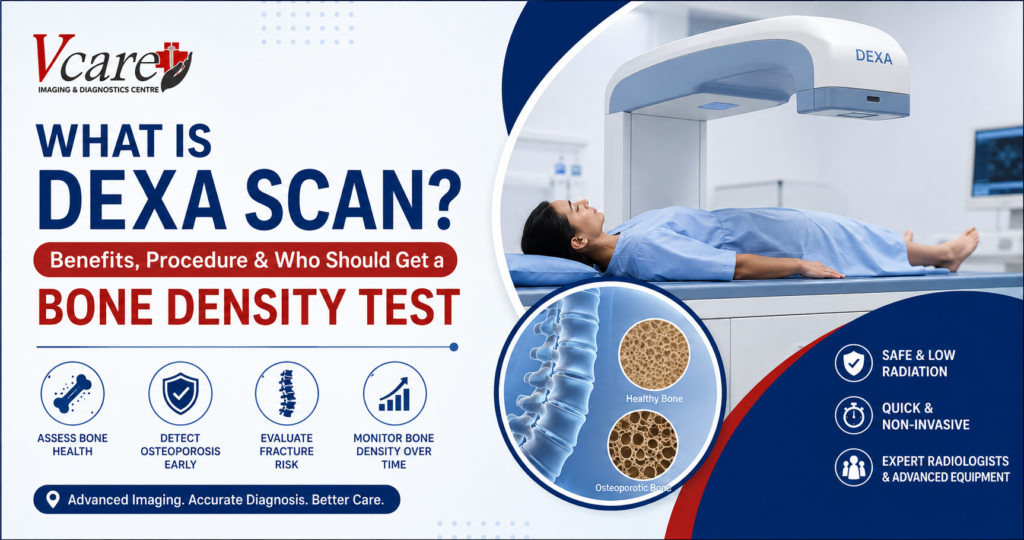
What Is DEXA Scan? Benefits, Procedure & Who Should Get a Bone Density Test

What Is a DEXA Scan? Benefits, Procedure & Who Should Get a Bone Density Test A DEXA scan is a specialized imaging test used to measure bone mineral density and assess bone health. DEXA stands for Dual-Energy X-ray Absorptiometry and is also commonly called a DXA scan or bone density test. The test uses a very small amount of X-ray radiation to measure bone mineral density, usually at the hip and lower spine. It is widely used to help diagnose osteoporosis, assess fracture risk, and monitor changes in bone density over time. A bone density test can be particularly useful when there are concerns about weakened bones or an increased risk of osteoporosis. Your doctor may recommend a DEXA scan based on your age, medical history, previous fractures, medications, or other risk factors. If you have been advised to undergo a DEXA scan in Kandivali, understanding the procedure and what the results mean can help you feel more prepared. Why Is a Bone Density Test Important? Bones naturally change throughout life. Bone loss can become more significant with age and may increase the risk of fractures. Osteoporosis is a condition in which bones become weaker and more fragile, increasing the likelihood of fractures. Because bone loss can develop without obvious symptoms, a bone density test can help identify reduced bone mineral density before a serious fracture occurs. A DEXA scan can help healthcare professionals: A DEXA scan does not predict with certainty whether an individual will experience a fracture. Instead, the results are considered along with other clinical risk factors to help doctors assess bone health and decide on appropriate management. What Does a DEXA Scan Measure? A DEXA bone density scan measures bone mineral density, commonly abbreviated as BMD. During the test, low-dose X-rays are used to measure the amount of X-ray energy absorbed by bone and soft tissue. The information is processed to calculate bone density measurements. The most commonly examined areas are: The hip and spine are commonly used because these areas are important for assessing bone health and fracture risk. 7 Benefits of a DEXA Scan 1. Helps Measure Bone Mineral Density The primary purpose of a DEXA scan is to measure bone mineral density. This provides doctors with objective information about the strength and mineral content of bones and can help identify low bone density. 2. Helps Detect Osteoporosis A DEXA scan is one of the standard imaging methods used to diagnose osteoporosis. It can identify reduced bone density and help healthcare professionals assess whether further evaluation or treatment may be appropriate. Early identification can be valuable because osteoporosis may progress without noticeable symptoms until a fracture occurs. 3. Helps Assess Fracture Risk Low bone density is associated with an increased risk of osteoporotic fractures. DEXA results can be considered alongside factors such as age, previous fractures, family history, body weight, lifestyle factors, and certain medical conditions when assessing fracture risk. 4. Supports Monitoring of Bone Health A bone density test may also be used to monitor changes in bone density over time. For people receiving treatment for osteoporosis or other conditions associated with bone loss, follow-up DEXA scans may help doctors evaluate changes and treatment response. 5. Quick and Non-Invasive A DEXA scan is generally a quick, non-invasive imaging examination. No anesthesia is normally required, and the patient typically lies on a padded examination table while the scanner obtains measurements. 6. Uses a Very Low Radiation Dose DEXA uses a small amount of ionizing radiation. The radiation exposure is considerably lower than that of many conventional X-ray examinations. Even so, always inform your doctor and the imaging technologist if you are pregnant or may be pregnant. 7. Helps Doctors Make Informed Decisions A DEXA scan provides measurable information about bone mineral density. When interpreted alongside your medical history and other risk factors, the results can help your healthcare provider make informed decisions about further evaluation, monitoring, or treatment. Who Should Get a DEXA Scan? Not everyone needs a DEXA scan routinely. The decision depends on individual risk factors and your doctor’s recommendation. A bone density test may be considered for people who have an increased risk of osteoporosis or bone loss. Your doctor may recommend a DEXA scan if you have: Postmenopausal women and older adults may have an increased risk of osteoporosis, but bone loss can also affect men and younger individuals in certain circumstances. The appropriate timing of a DEXA bone density scan should therefore be based on individual risk factors and medical advice. DEXA Scan for Women Bone density can decline after menopause, making bone health an important consideration for women as they age. A doctor may recommend a DEXA scan for women based on age, menopause status, fracture history, family history, medications, and other risk factors. Women who have additional risk factors for osteoporosis may need assessment earlier than someone without those risk factors. If you are concerned about bone health after menopause, discuss your individual risk factors with your doctor to determine whether a bone density test is appropriate. DEXA Scan for Men Osteoporosis is not limited to women. Men can also develop low bone density, particularly when certain medical conditions, medications, lifestyle factors, or age-related changes increase their risk. A doctor may recommend a DEXA scan for men when there are specific risk factors for osteoporosis or bone loss. What Happens During a DEXA Scan Procedure? Understanding the DEXA scan procedure can make the experience easier for first-time patients. Step 1: Patient Preparation Before the examination, the imaging team may ask about your medical history, medications, previous imaging examinations, and any relevant health conditions. You should tell your doctor or technologist if you may be pregnant. Step 2: Positioning You will usually lie on a padded examination table. For a central DEXA examination, the scanner generally evaluates the hip and lower spine. Your position may be adjusted so that the required areas can be accurately assessed. Step 3: Scanning The DEXA machine passes a scanning device over

What Is DEXA Scan? Benefits, Procedure & Who Should Get a Bone Density Test Read More »

breast ultrasound during breastfeeding

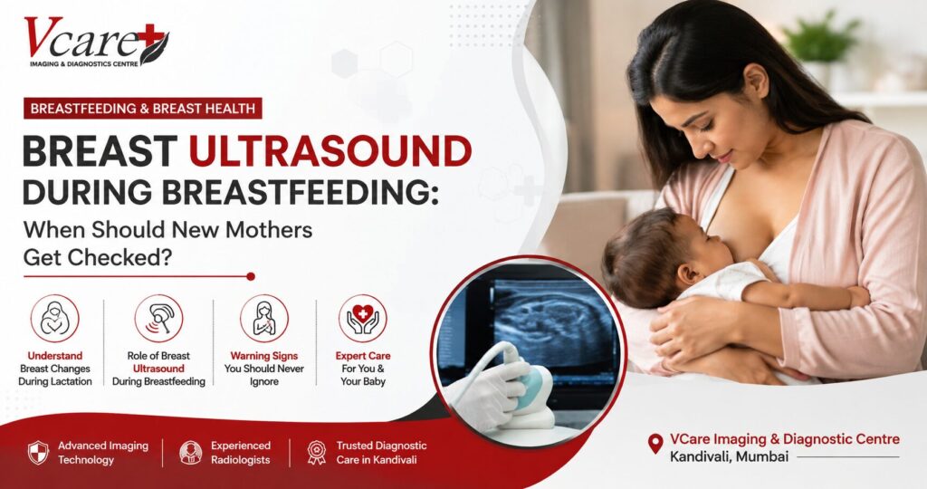
Breastfeeding & Breast Health: When Should a New Mother Consider a Breast Ultrasound?

Breastfeeding & Breast Health: When Should a New Mother Consider a Breast Ultrasound? Breastfeeding is a special phase of motherhood, but it also brings many physical changes. During lactation, the breasts naturally become fuller, denser, and more active as they produce milk. New mothers may occasionally notice tenderness, swelling, changes in breast texture, or even a lump. Although many breast changes during breastfeeding are benign, a new or persistent breast symptom should not simply be ignored because you are breastfeeding. A breast ultrasound during breastfeeding can be an important diagnostic tool when clinically recommended. It can help doctors evaluate a specific breast concern without using ionising radiation. At VCare Imaging & Diagnostic Centre in Kandivali, Mumbai, breast imaging services help women receive appropriate diagnostic evaluation based on their symptoms and their doctor’s or radiologist’s recommendations. Understanding what is normal during breastfeeding—and what deserves medical attention—can help new mothers make informed decisions about their breast health. Why Do Breasts Change During Breastfeeding? Pregnancy and breastfeeding cause significant hormonal and structural changes within breast tissue. Milk-producing glands become active, blood flow increases, and breasts may become larger and denser. These changes can sometimes make physical examination and breast imaging more complex. However, breastfeeding itself is not a reason to postpone evaluation of a concerning breast symptom. Medical imaging can be performed during lactation when clinically indicated. Some changes a breastfeeding mother may experience include: Many of these changes can have benign explanations. However, the cause of a persistent or unusual symptom cannot always be determined from symptoms alone. That is where appropriate clinical examination and, when recommended, breast imaging during breastfeeding can become important. What Is a Breast Ultrasound? A breast ultrasound, also known as breast sonography, uses high-frequency sound waves to create images of structures inside the breast. Unlike an X-ray, ultrasound does not use ionising radiation. During the examination, ultrasound gel is placed on the skin and a handheld transducer is moved over the area being examined. Images are produced in real time and interpreted by the appropriate medical professional. A breast ultrasound during breastfeeding may be used as part of the evaluation of a palpable lump, focal symptoms, or another breast concern, depending on the patient’s age, clinical findings and medical history. Current breast-imaging recommendations emphasize choosing imaging according to the specific clinical scenario rather than delaying investigation solely because a woman is lactating. When Should a Breastfeeding Mother Consider a Breast Ultrasound? Not every breastfeeding mother requires an ultrasound. Breast imaging is generally performed when there is a clinical reason or when recommended as part of an appropriate screening or diagnostic pathway. Here are some situations where a doctor may recommend further assessment. 1. A New or Persistent Breast Lump Finding a breast lump during breastfeeding can understandably cause anxiety. Lactation can be associated with benign conditions that may present as a lump. However, it is important not to assume that every lump is simply caused by breastfeeding. Current ACR guidance states that a new palpable abnormality during lactation should prompt appropriate imaging assessment. If you discover a lump that persists, changes, increases in size, or otherwise concerns you, consult your doctor rather than trying to diagnose it yourself. A breast ultrasound during breastfeeding may then form part of the diagnostic evaluation. 2. Persistent Localised Breast Pain Breast discomfort can occur during breastfeeding for several reasons. Feeding position, engorgement and other lactation-related issues may contribute to discomfort. However, persistent or focal breast pain deserves attention, particularly when accompanied by another symptom such as a lump, skin change, swelling or nipple discharge. A doctor can assess the symptoms and determine whether breast imaging is appropriate. 3. Swelling That Does Not Resolve Temporary fullness is common during breastfeeding. Persistent swelling—especially when confined to one area—should be discussed with a healthcare professional. Clinical evaluation helps determine whether the change appears related to breastfeeding or whether further assessment such as breast ultrasound is needed. 4. Persistent Signs of Infection or Inflammation Mastitis can occur during breastfeeding and may cause symptoms such as breast pain, redness, warmth and swelling, sometimes accompanied by fever or feeling unwell. Symptoms that do not improve as expected require medical review. Current ACR guidance specifically identifies unresolving infectious symptoms during lactation as a reason for imaging assessment. Ultrasound may be used in certain clinical situations to help assess whether there is a collection or another finding requiring further management. 5. Unusual Nipple Discharge Milk production is expected during breastfeeding, but a new or clinically suspicious nipple discharge should be evaluated rather than automatically attributed to lactation. The appropriate assessment depends on the nature of the discharge and other symptoms. ACR guidance includes clinically suspicious nipple discharge among the concerns that warrant imaging assessment during lactation. 6. Changes in the Skin or Nipple New breast changes should be discussed with a healthcare professional, particularly if they persist. These can include noticeable changes involving: These signs do not automatically indicate breast cancer, and there are many possible causes. The important point is that persistent or unexplained changes deserve proper evaluation. Is Breast Ultrasound Safe During Breastfeeding? Breast ultrasound does not involve ionising radiation and is widely used as a breast-imaging modality. The appropriate test, however, depends on factors including your symptoms, age, breast cancer risk and clinical examination. Women should therefore avoid assuming that ultrasound is always the only test needed simply because they are breastfeeding. Current evidence supports the use of ultrasound, mammography and, in selected circumstances, MRI for evaluating breast concerns in lactating patients. Can You Have a Mammogram While Breastfeeding? Yes. Mammography is not automatically contraindicated simply because you are breastfeeding. Current guidance supports mammography during lactation when it is appropriate for the patient’s age, risk or clinical situation. For example, the latest ACR criteria list mammography and digital breast tomosynthesis as usually appropriate for average-risk breast cancer screening in lactating women aged 40 or older. For diagnostic concerns, the imaging pathway may involve ultrasound, mammography, or both depending on age and the nature of the

Breastfeeding & Breast Health: When Should a New Mother Consider a Breast Ultrasound? Read More »

Best Time to Get a Mammography Test

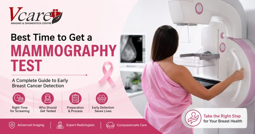
Best Time to Get a Mammography Test: A Complete Guide to Early Breast Cancer Detection

Best Time to Get a Mammography Test: Everything You Need to Know Breast cancer is one of the most common cancers affecting women worldwide. The good news is that early detection through regular mammography screening significantly increases the chances of successful treatment. Many women often wonder, “What is the best time to get a mammography test?” Understanding the right age, timing, and preparation for this screening can help detect abnormalities before symptoms even appear. At VCare Imaging & Diagnosis Center, we believe that awareness and timely screening play a vital role in maintaining breast health. This comprehensive guide explains when you should schedule your mammography, factors that influence the timing, preparation tips, and frequently asked questions. What Is a Mammography Test? A mammography test is a specialized X-ray examination of the breast designed to detect early signs of breast cancer and other breast abnormalities. It can identify tiny lumps or calcium deposits that may not be noticeable during a physical examination. Mammograms are considered the gold standard for breast cancer screening because they can detect cancer years before it becomes physically noticeable. Why Is Mammography Important? Early-stage breast cancer often develops without any obvious symptoms. A routine mammogram can detect changes in breast tissue long before a lump becomes large enough to feel. Benefits of Mammography Early detection often means less invasive treatment and better long-term outcomes. Best Time to Get a Mammography Test The best time to get a mammography test depends on several factors, including your age, menstrual cycle, family history, and overall risk of breast cancer. If You Are Between 40 and 49 Years Women in this age group should discuss screening recommendations with their healthcare provider. Depending on individual risk factors, mammography may be recommended every one to two years. If You Are 50 Years and Above Most healthcare professionals recommend getting a mammogram every one to two years. Regular screening during this age group plays a crucial role in detecting breast cancer early. If You Have a Family History of Breast Cancer Women with a strong family history or genetic mutations such as BRCA1 or BRCA2 may need to begin mammography screening before the age of 40. Your doctor may also recommend breast MRI in addition to mammography. Best Time During Your Menstrual Cycle If you are menstruating, scheduling your mammogram at the right time can improve comfort and image quality. The ideal time is: Avoid scheduling your mammogram just before your period because breast tenderness can make the examination more uncomfortable. Signs That You Should Not Delay a Mammography Even if your routine screening is not yet due, consult your doctor immediately if you notice: Diagnostic mammography may be recommended instead of routine screening. How Often Should You Get a Mammogram? The screening interval depends on your age and health history. Age Group Recommended Frequency Under 40 Only if recommended by your doctor 40–49 Every 1–2 years (based on risk) 50–74 Every 1–2 years Above 75 As advised by your healthcare provider How to Prepare for a Mammography Test Preparing properly ensures accurate imaging results. Before Your Appointment These simple steps help improve image quality and reduce the need for repeat scans. What Happens During the Procedure? A mammography examination usually takes around 15 to 30 minutes. During the procedure: Compression may feel uncomfortable for a few seconds, but it is necessary to obtain clear images while minimizing radiation exposure. Is Mammography Painful? Most women describe mammography as mildly uncomfortable rather than painful. The amount of discomfort varies depending on: Scheduling your appointment after your menstrual period often makes the examination more comfortable. Can Mammography Detect Breast Cancer Early? Yes. Mammography can detect breast cancer before symptoms appear, often identifying tumors that are too small to be felt during a self-examination. Studies have consistently shown that regular mammography screening reduces deaths from breast cancer by enabling earlier diagnosis and treatment. Who Is at Higher Risk? Certain women may require earlier or more frequent screening. Risk factors include: If you have one or more of these risk factors, consult your healthcare provider about a personalized screening schedule. Benefits of Regular Mammography Screening Routine mammograms offer several long-term health benefits. Early Detection Detects cancer before symptoms develop. Better Treatment Outcomes Earlier diagnosis often leads to less invasive treatment options and improved survival rates. Reduced Healthcare Costs Treating cancer at an early stage is generally less complex and less expensive than treating advanced disease. Improved Peace of Mind Regular screening helps women stay proactive about their breast health. Common Myths About Mammography Myth 1: Mammograms Cause Cancer False. Mammography uses a very low dose of radiation, and the benefits of early detection far outweigh the minimal risk. Myth 2: Only Women with Symptoms Need Mammograms False. Many breast cancers have no symptoms in their early stages, which is why routine screening is essential. Myth 3: Breast Self-Exams Replace Mammography False. Breast self-examinations are helpful for becoming familiar with your breasts, but they do not replace mammography. Imaging can detect abnormalities long before they can be felt. Frequently Asked Questions Why Choose VCare Imaging & Diagnosis Center? At VCare Imaging & Diagnosis Center, we are committed to providing accurate, comfortable, and patient-focused diagnostic services. Our advantages include: Our goal is to help patients receive accurate diagnoses while ensuring a smooth and reassuring experience. Final Thoughts Knowing the best time to get a mammography test is an important step toward protecting your breast health. Whether you are scheduling your first screening or maintaining a regular screening routine, timely mammography can help detect breast cancer at its earliest and most treatable stage. If you are over 40, have a family history of breast cancer, or notice any unusual breast changes, don’t delay your screening. Early detection saves lives, and regular mammograms remain one of the most effective tools for maintaining long-term breast health. Book your mammography appointment at VCare Imaging & Diagnosis Center today and take a proactive step toward early breast cancer detection and better health. https://maps.app.goo.gl/m8uoy6bBcuibEtkYA

Best Time to Get a Mammography Test: A Complete Guide to Early Breast Cancer Detection Read More »

Best 5 Diagnostic Centers in Kandivali (2026)

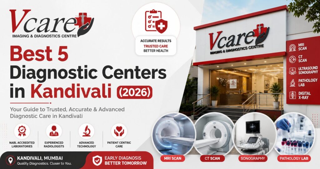
Best 5 Diagnostic Centers in Kandivali (2026) Finding the best diagnostic center in Kandivali is one of the most important decisions you can make for your health. Whether your doctor has recommended an MRI scan, CT scan, ultrasound, blood test, mammography, or a complete preventive health check-up, the quality of the diagnostic centre directly impacts the accuracy of your diagnosis and, ultimately, your treatment. A diagnostic test is more than just a report—it is the foundation of medical decision-making. Accurate imaging and pathology results help doctors identify diseases early, monitor existing conditions, and recommend the most effective treatment plan. Even the most experienced physician relies on high-quality diagnostic reports to provide the best possible care. Kandivali has several diagnostic centres offering imaging and laboratory services. However, the quality of technology, radiologists, reporting standards, hygiene, and patient care can vary significantly from one centre to another. Choosing a trusted diagnostic centre ensures you receive accurate results, timely reports, and a comfortable experience. To help you make an informed decision, we’ve compiled a list of some of the best diagnostic centers in Kandivali based on the range of services, advanced technology, patient experience, qualified specialists, and commitment to quality healthcare. Why Choosing the Right Diagnostic Centre Matters Many people select a diagnostic centre simply because it is close to home or offers low prices. While convenience and affordability are important, they should never come at the expense of diagnostic accuracy. A reliable diagnostic centre offers much more than routine testing. It combines experienced radiologists, modern imaging equipment, standardized protocols, skilled technicians, and quality control measures to ensure every report is accurate and clinically useful. Here are some key factors to consider when choosing a diagnostic centre: 1. Advanced Diagnostic Technology Modern equipment provides clearer images, faster scanning times, and greater diagnostic precision. Technologies such as Digital MRI, Multi-Slice CT Scan, Digital Mammography, 3D/4D Ultrasound, Colour Doppler, and Digital X-Ray improve image quality while enhancing patient comfort. 2. Experienced Radiologists Medical imaging requires expert interpretation. Qualified radiologists play a vital role in identifying abnormalities accurately, ensuring that reports are reliable and clinically meaningful. 3. Comprehensive Services Under One Roof A diagnostic centre that offers MRI, CT Scan, Ultrasound, Pathology, ECG, 2D Echo, Mammography, Bone Density Scan, and preventive health packages saves patients time and simplifies the diagnostic process. 4. Quality Standards Centres that follow recognized quality protocols and maintain high standards of hygiene, equipment maintenance, and reporting are generally more dependable. 5. Timely Reporting Quick and accurate reports help doctors begin treatment sooner, especially in emergency situations where time is critical. 6. Patient-Centric Care Friendly staff, clean facilities, efficient appointment scheduling, and clear communication all contribute to a better healthcare experience. 1. VCare Imaging & Diagnosis Center – Our Featured Recommendation When comparing the best diagnostic centers in Kandivali, VCare Imaging & Diagnosis Center stands out as a comprehensive destination for advanced imaging, pathology, and preventive healthcare services. With its focus on diagnostic excellence, modern technology, and patient-first approach, VCare has earned the trust of patients and referring doctors alike. Whether you need a routine blood test or an advanced MRI scan in Kandivali, VCare offers a wide range of diagnostic services under one roof, making healthcare more convenient and efficient. Comprehensive Diagnostic Services VCare provides an extensive range of diagnostic solutions, including: This comprehensive approach allows patients to complete multiple investigations at a single location, saving valuable time and reducing the need to visit multiple facilities. Advanced Imaging Technology Accurate diagnosis begins with high-quality imaging. VCare uses advanced diagnostic equipment designed to deliver detailed images that support precise medical evaluation. High-resolution imaging enables doctors to identify conditions affecting the brain, spine, joints, heart, lungs, abdomen, bones, and other organs with greater confidence. Modern technology also helps improve patient comfort by reducing scan times and producing clearer diagnostic images. Experienced Radiologists and Skilled Professionals Behind every accurate diagnostic report is an experienced team of radiologists and trained technicians. VCare’s medical professionals follow standardized imaging protocols and work closely with referring physicians to ensure every report provides meaningful clinical information. Their expertise plays a vital role in detecting diseases at an early stage and assisting doctors in making informed treatment decisions. Focus on Preventive Healthcare Healthcare today is not only about treating diseases-it is also about preventing them. VCare encourages regular preventive health screenings to identify potential health concerns before symptoms appear. Preventive health packages help monitor: Early diagnosis often leads to better treatment outcomes and lower healthcare costs. Comfortable Patient Experience Many patients feel anxious before undergoing MRI scans or other imaging procedures. VCare strives to create a welcoming environment where patients feel informed, supported, and comfortable throughout their visit. From appointment scheduling to report collection, the focus remains on delivering a smooth and hassle-free experience. Patients also appreciate: Why We Recommend VCare Imaging & Diagnosis Center While several diagnostic centres serve Kandivali, VCare stands out because of its combination of comprehensive services, advanced technology, experienced professionals, and patient-focused care. Key strengths include: For patients seeking reliable diagnostics, VCare offers a balanced combination of quality, convenience, and comprehensive healthcare services. Services Available at VCare Imaging & Diagnosis Center Who Should Visit VCare? VCare serves a wide range of patients, including: 2. Dr. Vyas Diagnostic Centre – A Well-Known Local Diagnostic Facility Dr. Vyas Diagnostic Centre is one of the established diagnostic centres serving patients in and around Kandivali. The centre offers a range of routine diagnostic services, including pathology tests, ultrasound, X-ray, and general health investigations. Many patients visit this centre for routine laboratory tests and basic diagnostic requirements. It caters to individuals looking for common health screenings and physician-recommended investigations. Key Services Dr. Vyas Diagnostic Centre is suitable for routine diagnostics. However, patients requiring advanced imaging modalities such as MRI, CT Scan, Digital Mammography, CBCT, or DEXA Scan may need to consider a centre with a broader range of diagnostic services. 3. Siddhi Diagnostic Centre Siddhi Diagnostic Centre has been serving local residents by providing pathology services and basic imaging investigations.

Best 5 Diagnostic Centers in Kandivali (2026) Read More »

Fatty Liver Diagnosis

Understanding Fatty Liver: Which Tests Help in Early Detection?

Understanding Fatty Liver: Which Tests Help in Early Detection? Fatty liver disease has become one of the most common lifestyle-related health conditions worldwide. It often develops silently, with little or no noticeable symptoms during its early stages. Because of this, many people remain unaware of the condition until it progresses into more serious liver damage. An accurate fatty liver diagnosis plays a vital role in detecting the condition early, allowing timely treatment and lifestyle modifications that can help prevent complications such as liver fibrosis, cirrhosis, and even liver failure. This guide explains everything you need to know about fatty liver diagnosis, including symptoms, causes, diagnostic tests, and when you should consider getting screened. What Is Fatty Liver Disease? Fatty liver disease occurs when excess fat accumulates inside the liver cells. While a small amount of fat is normal, excessive fat can affect liver function and increase the risk of inflammation and long-term liver damage. There are two primary types of fatty liver disease: Among these, NAFLD is becoming increasingly common due to unhealthy eating habits, obesity, diabetes, and sedentary lifestyles. Common Symptoms of Fatty Liver In many cases, fatty liver disease does not cause noticeable symptoms. However, as the condition progresses, patients may experience: Since these symptoms are often mild or absent, fatty liver diagnosis through regular health screenings becomes essential. Who Is at Risk? Certain individuals have a higher risk of developing fatty liver disease. You may be at higher risk if you have: Routine fatty liver diagnosis is recommended for individuals with these risk factors. Which Tests Help in Fatty Liver Diagnosis? Several diagnostic tests help doctors evaluate liver health and determine the severity of fatty liver disease. 1. Liver Function Test (LFT) A Liver Function Test measures enzymes and proteins in your blood that indicate how well your liver is functioning. Although an LFT cannot confirm fatty liver by itself, abnormal liver enzyme levels often prompt further investigations. 2. Ultrasound Scan Ultrasound is one of the most commonly recommended imaging tests for fatty liver diagnosis. It is: An abdominal ultrasound can identify fat deposits inside the liver before symptoms become severe. 3. FibroScan (Liver Elastography) FibroScan is an advanced, non-invasive test that measures liver stiffness and fat content. Doctors recommend FibroScan to: 4. CT Scan A CT scan provides detailed cross-sectional images of the liver and surrounding organs. While not always the first choice for fatty liver diagnosis, it can help detect liver abnormalities when additional imaging is required. 5. MRI Scan MRI offers highly detailed liver imaging and is particularly useful for accurately measuring liver fat content. It is often recommended in complex cases or when precise evaluation is needed. 6. Blood Tests Doctors may also recommend additional blood investigations, including: These tests help identify underlying conditions contributing to fatty liver disease. Why Early Fatty Liver Diagnosis Matters Early detection significantly improves treatment outcomes. Benefits include: The earlier the fatty liver diagnosis, the easier it is to manage the condition. Can Fatty Liver Be Reversed? Yes. In many cases, early-stage fatty liver disease is reversible. Doctors usually recommend: Regular follow-up testing helps monitor liver health and treatment progress. When Should You Get Screened? You should consider a fatty liver diagnosis if you: Routine health screenings can detect fatty liver before serious complications develop. Why Choose VCare Imaging & Diagnostic Centre? At VCare Imaging & Diagnostic Centre, we provide advanced diagnostic services to support accurate fatty liver diagnosis. Our services include: Our goal is to help patients detect liver conditions early for timely treatment and improved health outcomes. Conclusion Fatty liver disease often develops silently, making regular health screenings essential for early detection. An accurate fatty liver diagnosis using imaging tests and blood investigations allows doctors to identify the condition before permanent liver damage occurs. If you have risk factors such as obesity, diabetes, or high cholesterol, don’t wait for symptoms to appear. Early diagnosis and timely lifestyle changes can protect your liver and improve your overall well-being. Schedule your liver screening today and take the first step toward better liver health. Frequently Asked Questions (FAQs)

Understanding Fatty Liver: Which Tests Help in Early Detection? Read More »

Preventive Health Checkup

Preventive Health Checkups: Why Early Detection Is the Key to a Healthier Life

Preventive Health Checkups: Why Early Detection Is the Key to a Healthier Life In today’s fast-paced world, many people focus on treating illnesses only after symptoms appear. However, by the time noticeable symptoms develop, a health condition may already have progressed. This is why a preventive health checkup plays such a vital role in maintaining long-term health and well-being. Preventive healthcare focuses on identifying potential health issues before they become serious. Through routine blood tests, diagnostic imaging, and comprehensive health screenings, doctors can detect diseases at an early stage, allowing for timely treatment and better outcomes. Whether you are a young professional, a busy parent, or a senior citizen, scheduling a preventive health checkup is one of the smartest investments you can make for your future health. What Is a Preventive Health Checkup? A preventive health checkup is a comprehensive medical evaluation designed to assess your overall health and identify potential health risks before symptoms appear. Unlike diagnostic tests, which are performed to investigate existing symptoms, preventive health screenings help detect diseases in their earliest stages. These checkups typically include: Depending on your age, lifestyle, family history, and medical condition, your doctor may recommend additional diagnostic tests or imaging studies. Why Early Detection Matters One of the greatest advantages of a preventive health checkup is early disease detection. Many serious medical conditions develop silently without causing obvious symptoms. Early diagnosis can significantly improve treatment outcomes for conditions such as: When these conditions are identified early, treatment is often simpler, less expensive, and more effective. Regular health screenings also help prevent complications and reduce the risk of hospitalization. Benefits of Preventive Health Checkups 1. Detect Diseases Before Symptoms Appear Many chronic diseases remain unnoticed for months or even years. Routine diagnostic tests help uncover hidden health problems before they become serious. 2. Reduce Long-Term Healthcare Costs Preventing disease is generally more affordable than treating advanced medical conditions. Early intervention can reduce hospitalization, surgery, and long-term medication costs. 3. Monitor Existing Health Conditions Individuals with diabetes, hypertension, thyroid disorders, or heart disease benefit from regular monitoring through laboratory investigations and imaging studies. 4. Improve Overall Quality of Life Knowing your health status allows you to make informed lifestyle changes, including improving your diet, increasing physical activity, managing stress, and maintaining a healthy weight. 5. Peace of Mind Regular health screenings provide reassurance when results are normal and help identify problems early when treatment is most effective. Who Should Get a Preventive Health Checkup? A preventive health checkup is beneficial for adults of all ages, but certain groups may benefit even more from routine screenings. Adults Above 30 As the risk of lifestyle-related diseases increases with age, annual health checkups become increasingly important. Working Professionals Long working hours, stress, sedentary lifestyles, poor eating habits, and lack of exercise increase the risk of chronic illnesses. Senior Citizens Older adults should undergo regular screenings for heart health, bone health, kidney function, vision, and other age-related conditions. Individuals with a Family History of Disease If your family has a history of diabetes, hypertension, heart disease, thyroid disorders, or cancer, preventive screening helps assess your risk and supports early intervention. Common Diagnostic Tests Included in Preventive Health Checkups A comprehensive preventive health checkup may include a combination of laboratory tests and diagnostic imaging based on your health profile. Blood Tests Routine blood investigations evaluate: Blood Pressure Monitoring Regular blood pressure checks help identify hypertension before complications develop. ECG An Electrocardiogram evaluates heart rhythm and may detect early signs of cardiovascular disease. Chest X-Ray A chest X-ray helps assess lung health and identify infections or abnormalities when clinically indicated. Ultrasound Ultrasound imaging provides valuable information about abdominal organs such as the liver, kidneys, gallbladder, pancreas, and reproductive organs. Bone Health Assessment Individuals at risk of osteoporosis may benefit from bone density evaluation as advised by their healthcare provider. How Often Should You Have a Preventive Health Checkup? The ideal frequency depends on several factors, including age, medical history, lifestyle, and existing health conditions. General recommendations include: Your healthcare provider can suggest a personalized screening schedule based on your individual health needs. Tips to Make the Most of Your Health Checkup To ensure accurate results: These simple steps help your healthcare team provide a more accurate assessment and personalized advice. Why Choose VCare Imaging & Diagnostic Centre? Choosing a trusted diagnostic centre is just as important as scheduling a preventive health checkup. At VCare Imaging & Diagnostic Centre, we are committed to delivering accurate, timely, and patient-focused diagnostic services. Our experienced team, advanced diagnostic technology, and comprehensive range of pathology and imaging services help support early disease detection and informed medical decisions. Whether you require routine blood investigations, ultrasound, X-ray, MRI, CT scan, or a complete health screening, we strive to provide reliable diagnostics in a comfortable environment. Regular preventive health checkups empower you to take control of your health before problems arise. Investing in routine screening today can help you enjoy a healthier tomorrow. Conclusion A preventive health checkup is one of the most effective ways to protect your long-term health. Early disease detection allows timely treatment, reduces complications, and supports a better quality of life. Rather than waiting for symptoms to appear, make preventive healthcare a regular part of your wellness routine. By combining healthy lifestyle habits with routine health screenings and diagnostic tests, you can take proactive steps toward a healthier future for yourself and your family. Frequently Asked Questions (FAQs)

Preventive Health Checkups: Why Early Detection Is the Key to a Healthier Life Read More »

Monsoon Health Screening for preventive healthcare

Monsoon Health Screening: Why Preventive Diagnostic Tests Matter More During the Rainy Season

Monsoon Health Screening: Why Preventive Diagnostic Tests Matter More During the Rainy Season The arrival of the monsoon season brings welcome relief from the summer heat, lush greenery, and refreshing rainfall. However, along with the pleasant weather comes an increased risk of infections, waterborne diseases, and seasonal health complications. From viral fevers and respiratory infections to dengue and malaria, monsoon-related illnesses can affect people of all ages. This is where Monsoon Health Screening becomes essential. A proactive approach to health through timely diagnostic tests can help identify potential health concerns before they become serious. Instead of waiting for symptoms to worsen, preventive health screenings allow individuals to stay informed about their health and take necessary precautions during the rainy season. In this blog, we’ll explore why a Monsoon Health Screening is important, the key diagnostic tests to consider, and how preventive healthcare can help you enjoy a healthier monsoon. Why Is Health Risk Higher During the Monsoon? The rainy season creates favorable conditions for the growth of bacteria, viruses, and mosquitoes. Stagnant water becomes a breeding ground for disease-carrying insects, while increased humidity can contribute to respiratory and fungal infections. Common health concerns during monsoon include: Many of these conditions initially present with mild symptoms that can easily be overlooked. Early diagnosis through a Monsoon Health Screening can help detect underlying infections and prevent complications. What Is a Monsoon Health Screening? A Monsoon Health Screening is a preventive health assessment designed to evaluate your overall health and identify any early signs of infection or illness commonly associated with the rainy season. These screenings typically include blood tests, urine tests, and other diagnostic evaluations that provide valuable insights into your health status. The goal is to detect abnormalities before they lead to severe health issues. Preventive healthcare is not only about treating illnesses but also about preventing them. By undergoing a Preventive Health Checkup during monsoon, individuals can make informed decisions regarding their health and lifestyle. Benefits of Monsoon Health Screening 1. Early Detection of Seasonal Diseases Many monsoon-related diseases begin with common symptoms such as fever, fatigue, headache, and body pain. Diagnostic tests help determine the exact cause and ensure timely treatment. Early detection can significantly reduce the risk of complications associated with diseases like dengue, malaria, and typhoid. 2. Monitoring Overall Health A Health Checkup in Monsoon does more than detect infections. It provides a comprehensive overview of important health parameters, including blood sugar levels, liver function, kidney function, and immune response. Regular monitoring helps individuals maintain optimal health throughout the season. 3. Preventing Serious Health Complications Ignoring early symptoms can lead to delayed diagnosis and prolonged recovery. Preventive screenings identify potential concerns before they become severe, allowing for prompt medical intervention. 4. Peace of Mind Knowing that your health parameters are within a healthy range offers reassurance and confidence during a season known for increased health risks.\ Essential Diagnostic Tests During Monsoon A comprehensive Monsoon Health Screening may include several important tests depending on individual health needs and risk factors. Complete Blood Count (CBC) A CBC test helps evaluate overall health and can detect infections, anemia, and abnormalities in blood cells. It is often one of the first tests recommended when experiencing fever or fatigue. Dengue Profile Test Dengue cases tend to rise during monsoon due to mosquito breeding. A dengue profile test helps identify the infection early and monitor platelet levels. Malaria Test Malaria remains a common concern during the rainy season. Early detection through diagnostic testing enables timely treatment and prevents complications. Typhoid Test Contaminated food and water can increase the risk of typhoid during monsoon. Testing helps confirm infection and guide appropriate medical care. Liver Function Test (LFT) Certain infections can affect liver health. Liver function tests assess how well the liver is working and help identify any abnormalities. Kidney Function Test (KFT) Kidney function tests evaluate the health of the kidneys and ensure they are functioning properly. Urine Analysis Urine testing can detect infections, dehydration, and other health conditions that may require medical attention. Chest X-Ray (If Recommended) For individuals experiencing persistent cough, breathing difficulties, or respiratory symptoms, a chest X-ray may help identify underlying lung conditions. Who Should Consider a Monsoon Health Screening? While everyone can benefit from preventive healthcare, certain groups should prioritize Diagnostic Tests During Monsoon: Children Children are more vulnerable to infections due to their developing immune systems. Senior Citizens Older adults often have weaker immunity and may be at higher risk of seasonal illnesses. Individuals with Chronic Conditions People with diabetes, hypertension, asthma, or heart disease should consider regular health monitoring during monsoon. Working Professionals Busy schedules often lead to delayed medical attention. Preventive screenings help identify concerns before they affect productivity and well-being. Tips to Stay Healthy During Monsoon Along with regular Monsoon Health Screening, adopting healthy habits can help reduce the risk of illness. Maintain Proper Hygiene Wash your hands frequently and avoid consuming unhygienic food. Drink Safe Water Always drink filtered or boiled water to reduce the risk of waterborne diseases. Eat Fresh and Nutritious Food A balanced diet rich in fruits, vegetables, and essential nutrients supports a strong immune system. Prevent Mosquito Breeding Remove stagnant water around your home and use mosquito repellents when necessary. Stay Physically Active Regular exercise helps strengthen immunity and improve overall health. Seek Medical Advice Early Do not ignore symptoms such as persistent fever, fatigue, or unusual discomfort. Early consultation and diagnostic testing can make a significant difference. Why Choose a Trusted Diagnostic Center for Monsoon Health Screening? The accuracy of diagnostic results plays a crucial role in healthcare decisions. Choosing a reliable diagnostic center ensures access to advanced technology, experienced healthcare professionals, and precise reporting. A trusted diagnostic center provides comprehensive screening services, enabling individuals and families to take proactive steps toward better health during the monsoon season. Whether you are experiencing symptoms or simply want to stay ahead of potential health risks, investing in a Monsoon Health Screening is a smart and responsible choice. Conclusion The monsoon

Monsoon Health Screening: Why Preventive Diagnostic Tests Matter More During the Rainy Season Read More »

Diagnostic Tests After Age 40

Top 5 Diagnostic Tests Everyone Should Consider After Age 40

Top 5 Diagnostic Tests After Age 40 Everyone Should Consider Turning 40 is an important milestone in life, and it is also the right time to take your health more seriously. As we age, the risk of developing conditions such as heart disease, diabetes, hypertension, osteoporosis, liver disorders, and certain cancers increases significantly. Regular diagnostic tests after age 40 can help detect health issues at an early stage, allowing timely treatment and better outcomes. Many serious diseases develop silently without noticeable symptoms. This is why preventive healthcare and routine health screenings are essential for maintaining long-term wellness. At VCare Imaging & Diagnostic Center, we believe that early diagnosis is the key to effective treatment and a healthier future. In this guide, we will discuss the top 5 diagnostic tests everyone should consider after age 40 and why these screenings are crucial for your overall health. Why Are Diagnostic Tests Important After Age 40? After the age of 40, the body’s metabolism begins to slow down, and the likelihood of chronic health conditions increases. Regular health checkups after 40 can help: Preventive healthcare is always more effective and affordable than treating advanced-stage illnesses. 1. Full Body Health Checkup A comprehensive full-body health checkup is one of the most important health screenings for individuals above 40. What Does It Include? Benefits Doctors generally recommend an annual health checkup after the age of 40 to maintain optimal health. 2. MRI Scan for Early Detection of Internal Conditions An MRI Scan uses advanced magnetic imaging technology to provide detailed images of organs, joints, soft tissues, and the brain. Why Is MRI Important After 40? As people age, they become more susceptible to: Common MRI Examinations MRI scans help doctors diagnose conditions accurately without exposing patients to radiation. 3. CT Scan for Comprehensive Internal Assessment A CT Scan combines multiple X-ray images to create detailed cross-sectional views of the body. Conditions Detected Through CT Scan Who Should Consider a CT Scan? Individuals with: A CT scan plays a vital role in early disease detection and treatment planning. 4. Mammography and Breast Screening (For Women) Breast cancer remains one of the most common cancers among women worldwide. Regular digital mammography can significantly improve the chances of early detection. Why Mammography Is Essential After 40 Healthcare experts recommend annual breast screening for women aged 40 and above, especially if they have: Benefits of Mammography Early detection can save lives and reduce the need for extensive treatment. 5. Ultrasound and Abdominal Screening An ultrasound scan is a safe, non-invasive diagnostic test that helps evaluate internal organs. What Can an Ultrasound Detect? Why Is It Recommended After 40? Lifestyle-related conditions such as obesity, diabetes, and fatty liver disease become more common with age. Regular ultrasound screening helps identify these problems before they become severe. Additional Health Screenings to Consider Besides the top five diagnostic tests after age 40, healthcare professionals may also recommend: Bone Density Scan To assess osteoporosis risk and bone health. Cardiac Screening Including ECG, 2D Echo, and stress tests for heart health evaluation. Blood Pressure Monitoring To detect hypertension and cardiovascular risks. Colon Cancer Screening Especially for individuals with a family history of colorectal cancer. Liver Elastography For evaluating liver stiffness and detecting liver fibrosis. Signs That Indicate You Need Diagnostic Testing Do not wait for symptoms to become severe. Consider diagnostic screening if you experience: Early diagnosis leads to faster treatment and better health outcomes. How Often Should You Get Tested After 40? The frequency of diagnostic tests depends on factors such as: In general: Diagnostic Test Recommended Frequency Full Body Checkup Once a Year Blood Sugar Test Every 6-12 Months MRI Scan As Recommended by Doctor CT Scan When Clinically Required Mammography Annually After 40 Ultrasound Every 1-2 Years Always consult your healthcare provider for personalized recommendations. Why Choose VCare Imaging & Diagnostic Center? At VCare Imaging & Diagnostic Center, we provide advanced imaging and diagnostic services using modern technology and expert medical professionals. Our services include: We are committed to delivering accurate reports, timely diagnosis, and exceptional patient care to help you make informed healthcare decisions. Conclusion Regular diagnostic tests after age 40 play a critical role in maintaining long-term health and preventing serious medical conditions. A proactive approach to health screening can help detect diseases early, improve treatment outcomes, and enhance your quality of life. Whether it is a full body health checkup, MRI scan, CT scan, mammography, or ultrasound screening, investing in preventive healthcare today can protect your health for years to come. Schedule your health screening with VCare Imaging & Diagnostic Center and take the first step toward a healthier future.

Top 5 Diagnostic Tests Everyone Should Consider After Age 40 Read More »

Scroll to Top